Learning Objectives
By the end of this section, you will be able to:
- Identify, locate, and describe the origin, insertion, and function of three major muscles in the head and neck region.
We will consider the following three muscles:
- Masseter
- Sternocleidomastoid
- Trapezius
Instructions:
You’ve learned the major bones in the last two labs—now it’s time to put that knowledge to work! Follow these steps as you explore each muscle:
- Observe the rotating GIFs for each muscle to visualize its position.
- Use the Innerbody 3D Model to explore each muscle in the context of the whole body and neighboring muscles.
- Activate the 3D function. Rotate the model and click on muscles to reveal their names.
- Try to locate the three muscles featured in this section.
- Tip: Use the selection tool (arrow) to hide superficial muscles and reveal the deeper layers.
- You can also toggle through the anatomy layers to reveal the bone origin and insertion sites of each muscle.
- Predict each muscle’s origin and insertion based on its position and name.
- Ask yourself: Which bones form the landmarks mentioned in this muscle’s name?
- Once you’ve made your best guess, open the dropdowns to check your understanding.
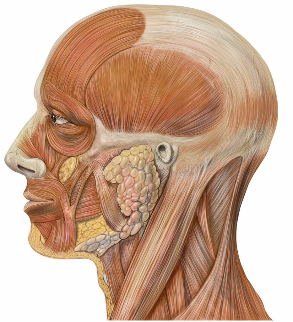
Masseter
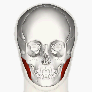
This cheek muscle has a superficial and a deep part and is named after its function.
Sternocleidomastoid
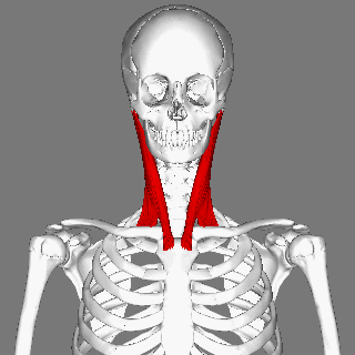
This long narrow muscle has two heads and is in the neck. It is named for two of its origin sites and its one insertion site. Can you see from the name and image what these sites might be?
🌐 Explore the sternocleidomastoid in 3D: Click here and use the rotation tools from UMich Anatomy to identify the origin and insertion points on a skeleton.
TRAPEZIUS
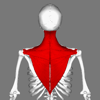
This is a large, flat, triangular sheet of muscle. It covers the posterior neck region and the upper region of the back. It attaches the axial skeleton to the upper appendicular skeleton. Can you see this on the diagram? Looking at the diagram, why do you think it’s named “trapezius”?
🌐Visit the AnatomyZone site. Move the image around in 3D to observe and make a note of this muscle’s sites of origin and insertion. Remember that you can click on the bones to recall their names. You can also consult the UMich Anatomy site for a slightly different view.
✍️Make a visual note on how this muscle connects the axial and appendicular skeletons. Based on this muscle’s multiple origin and insertion sites, can you guess what its function may be? See if you got this right using the dropdowns below.