Learning Objectives
By the end of this section, you will be able to:
Identify, locate, and describe the origin, insertion and function of four muscles of the shoulder and back.
We will consider the following four muscle groups:
- pectoralis major
- deltoid
- serratus anterior
- latissimus dorsi
Pectoralis major
The pectoral major may colloquially be referred to as “pecs” due to it being the largest and most superficial muscle in the chest area. The pectoralis major (from Latin pectus ‘breast’) is a thick, triangular and fan-shaped muscle that covers both sides of the upper region of the chest (only one side is shown in the figure above).
Deltoid

This is the major muscle of the lateral region of the glenohumeral joint (shoulder joint). It is named after the Greek letter delta, which means triangular. Its three fibres can contract independently or simultaneously.
clavicle, acromion process of scapula and from the scapular spine
Helpful mnemonic (KenHub):
Deltoid helps you carry SACS:
- Clavicle
- Acromion
- Spine of Scapula
deltoid tuberosity of humerus
Its primary functions is abduction of the arm along with other muscles. Due to its multiple origin sites, the deltoid can also rotate the arm.
As shown below, contraction of the deltoid pulls the head of the humerus into the glenoid cavity.
Serratus anterior
This broad muscle can be seen underlying the pectoralis muscles. It forms much of the lateral musculature of the axillary (underarm) region in the lateral wall of the thorax. Try to identify its origin and insertion sites on the figure. After identifying this muscle, can you think of the reason that it’s called ‘serratus’?
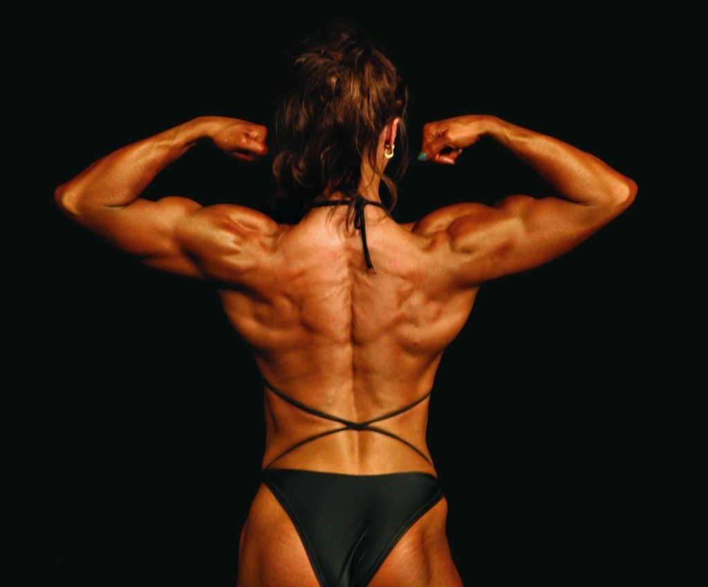
Latissimus dorsi
This large, flat muscle on the back stretches to the sides, behind the arm, and is partly covered by the trapezius on the back near the midline. The latissimus dorsi (AKA: ‘the lats muscle’ or ‘the lats’) is the widest muscle in the human body (KenHub). This muscle is named for the latin word that means “broadest” and dorsi for back.
Visit this AnatomyZone site. Click on ‘View Interactive 3D model’ below the video. Rotate the image in 3D using your mouse to view the origins and insertion. You can also click on the bones for identification.