
Learning Objectives
By the end of this section, you will be able to:
Identify, locate, and describe the origin, insertion and function of the muscles of the thorax.
The muscles of the chest serve to facilitate breathing by changing the size of the thoracic cavity. When you inhale, your chest rises because the cavity expands. Alternately, when you exhale, your chest falls because the thoracic cavity decreases in size.
Breathing involves two events: inspiration (inhalation), when the air moves into the lungs and expiration (exhalation), when the air leaves the lungs.
We will consider the following three muscles of the thorax:
- External intercostals
- Internal intercostals
- Diaphragm
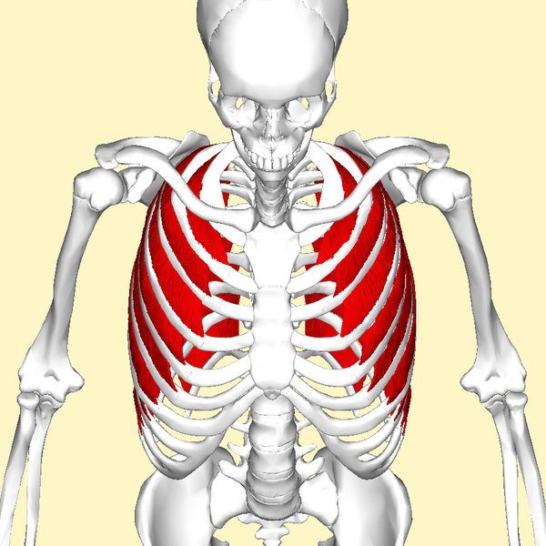
Fibers of these muscles slope downward and forward (toward the midline) (think hands in your pockEts). This means that these muscles are oriented inferiorly and medially. They originate from the lower border of one rib and insert on the upper border of the rib immediately inferior.
Function: to expand the chest cavity during inspiration (opposite of E).
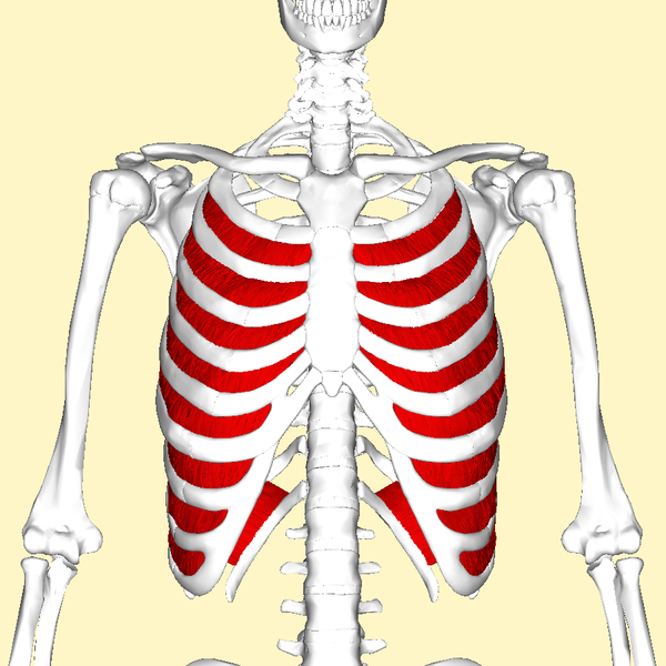
These muscles lie immediately deep to the external intercostals and their fibers run obliquely in the opposite direction. The internal intercostal muscles are oriented downwards and backwards away from the sternum (think ‘hands on your chIn’).
The internal intercostals originate on the costal grooves of the first to eleventh ribs and their adjacent costal cartilages. They insert on the superior borders of ribs and costal cartilages immediately below their origin sites.
Function: forced expiration (opposite it I). These muscles reduce the diameter of the thoracic cavity by depressing the ribs, thus pushing the air out of the lungs. This means that the internal intercostals are only used in forceful exhalation such as coughing or during exercise and not in relaxed breathing.
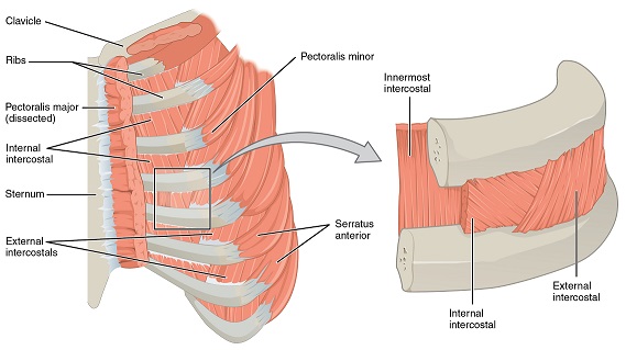
The relative positions and arrangement of the internal and external intercostal muscles can be seen in the figure below. The external intercostals are located laterally on the sides of the body. The internal intercostals are located medially near the sternum.
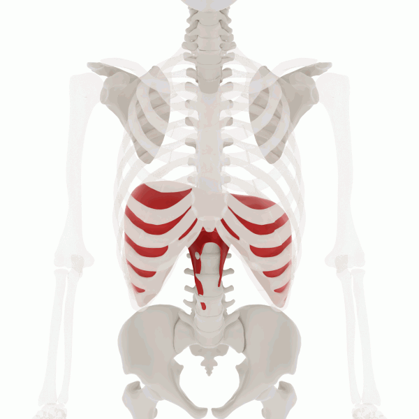
The most important muscle of inspiration, the broad diaphragm is a skeletal muscle that forms the floor of the thoracic cavity. In the relaxed state, the diaphragm is dome shaped, but when it contracts it moves inferiorly and flattens, increasing the volume of the thoracic cavity. This expands the volume of the cavity to allow the lungs to expand and fill with air. Therefore, the primary function of its contraction is inspiration. It also can be contracted voluntarily to increase pressure on the abdominal cavity to urinate, defecate or deliver a baby.
The gif below shows that when the diaphragm contracts, it moves inferiorly and flattens out to push out the thoracic cavity. 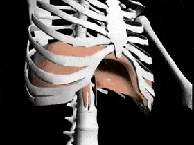
Watch the video below on the muscles of the thoracic wall (12:33 min).
Test Your Knowledge
Now it’s your turn! Use the interactive activities below to test your knowledge of muscles of the thorax (and shoulder).