Learning Objectives
By the end of this section, you will be able to:
Identify, locate, and describe the origin, insertion and function of the major upper arm and forearm muscles.
Muscles of the upper arm
We will first consider the following two muscle groups with opposing functions of flexion and extension:
- Biceps brachii
- Triceps brachii
Biceps brachii
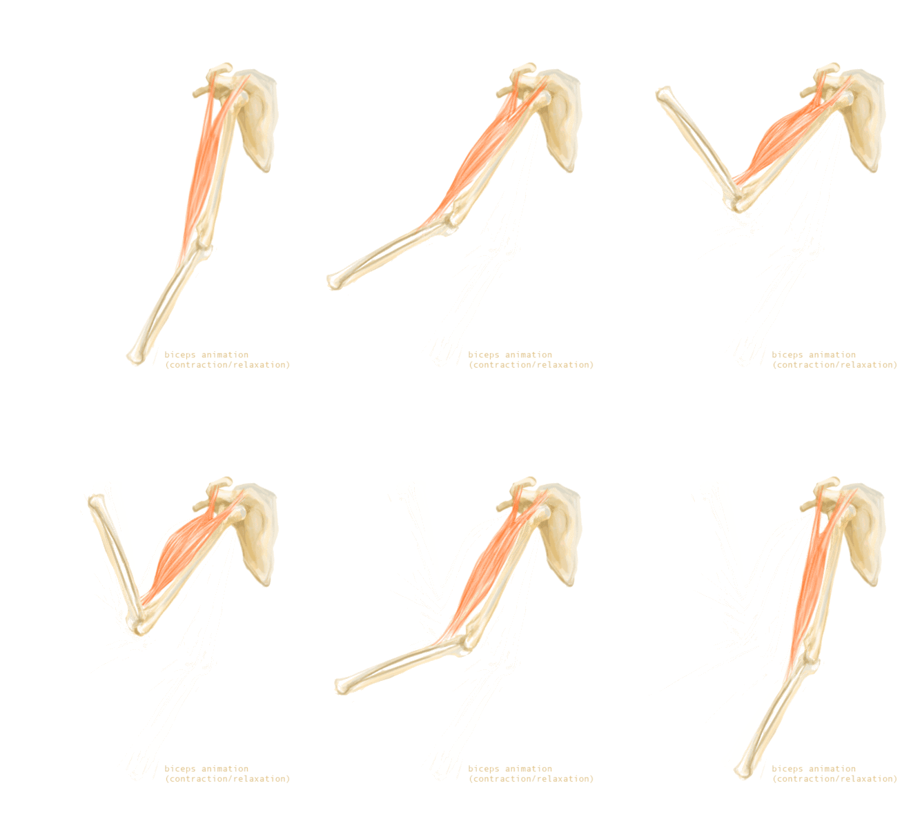
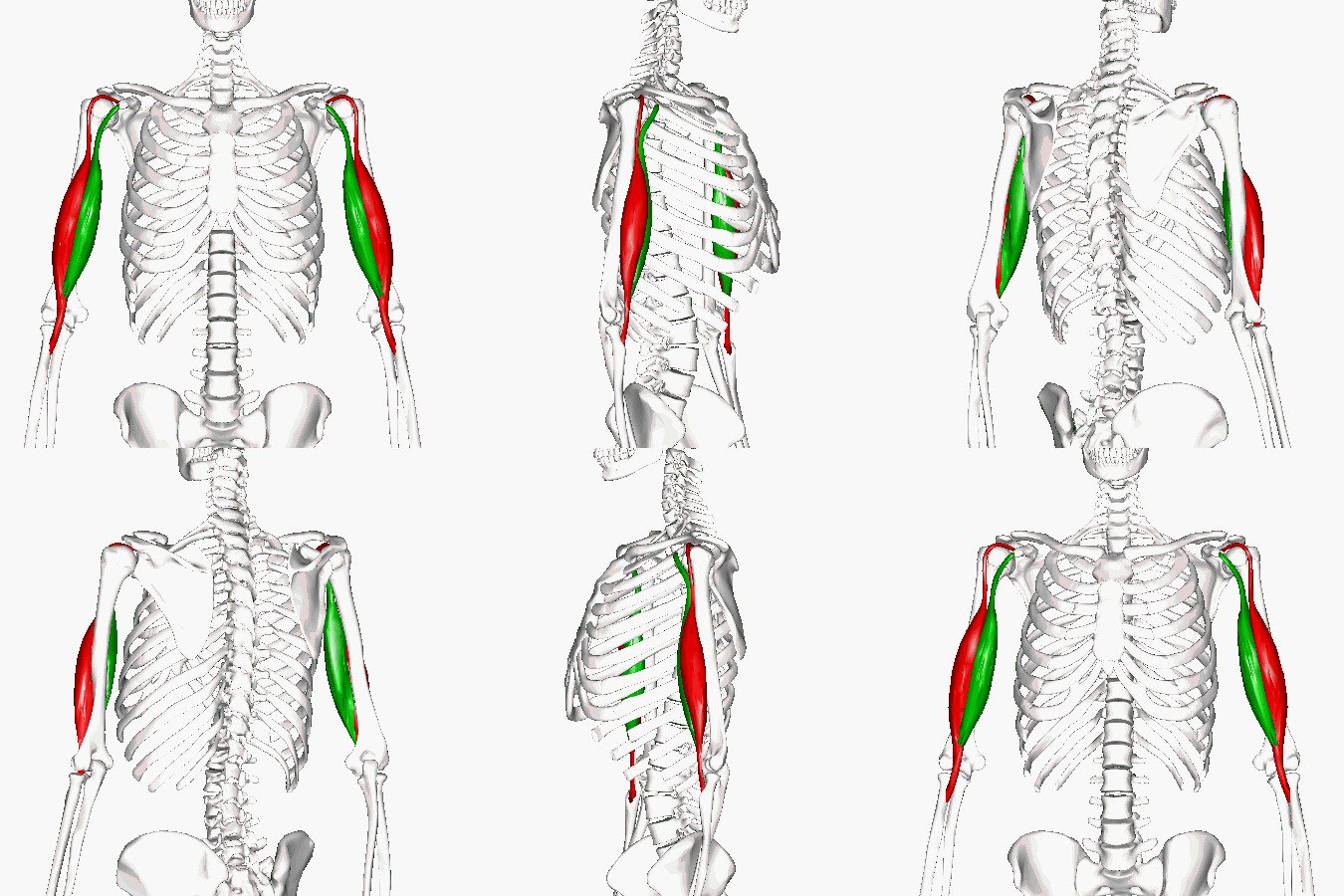
This is the large two-headed muscle covering the anterior of the humerus.
You can see the two heads in green and red on the humerus in the diagram to the right.
Visit this AnatomyZone site. Click on ‘View Interactive 3D model’ below the video. Rotate the image in 3D using your mouse to view the origins and insertion of the biceps brachii.
Triceps brachii
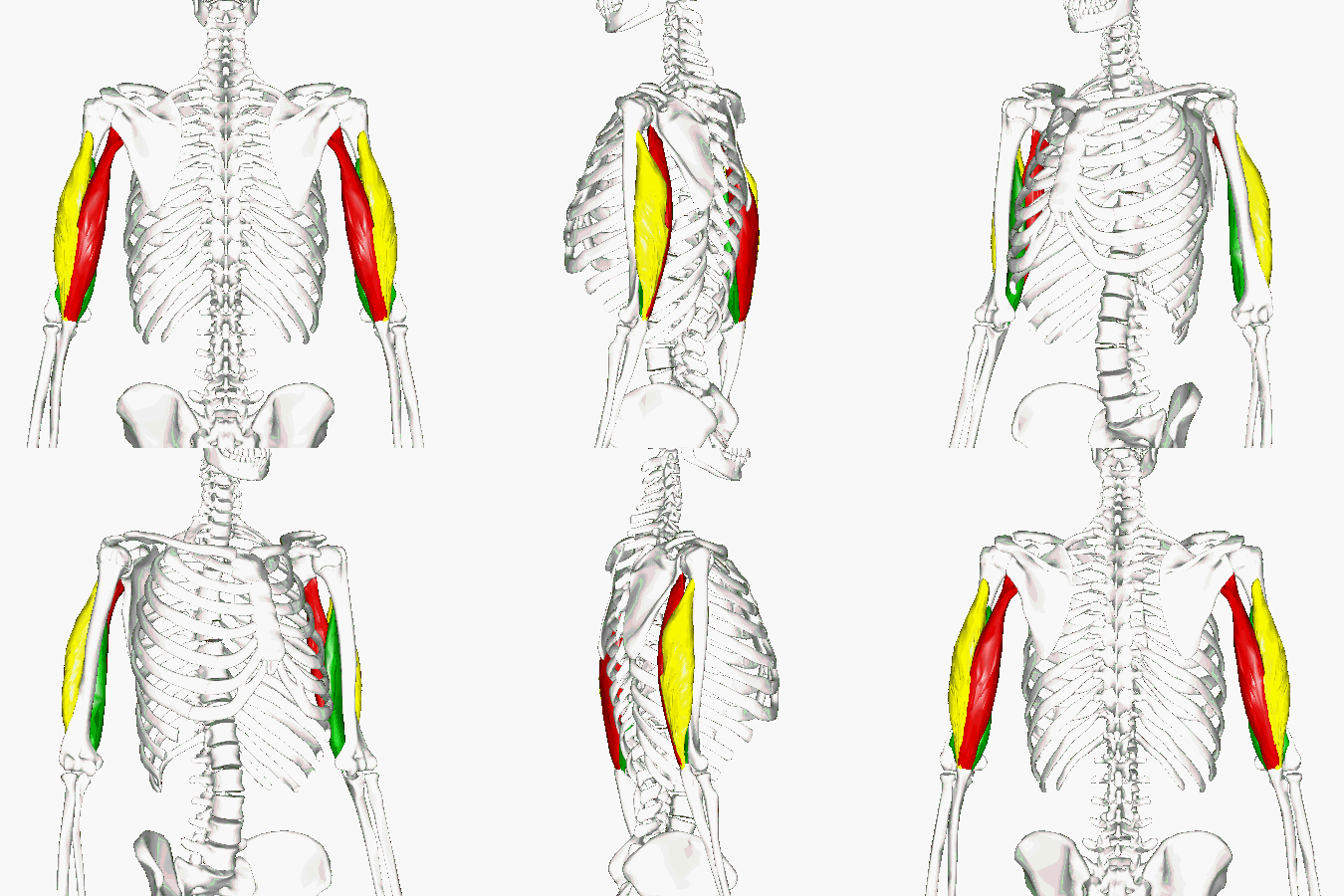
This three-headed muscle covers the posterior surface of the upper arm. Hint…it is not on the thumb side. Look at the image to the right and see if you can identify on which two bones the heads of this muscle originate?
Take another look at the image and try to locate the bone upon which this muscle inserts.
Visit this AnatomyZone site. Click on ‘View Interactive 3D model’ below the video. Rotate the image in 3D using your mouse to view the origins and insertion of the triceps brachii. Use the right/left arrows to scroll to page two for a description of the origins of the three heads and their insertion site. Click on each bone for identification and review.
Muscles of the forearm that don’t extend into the digits
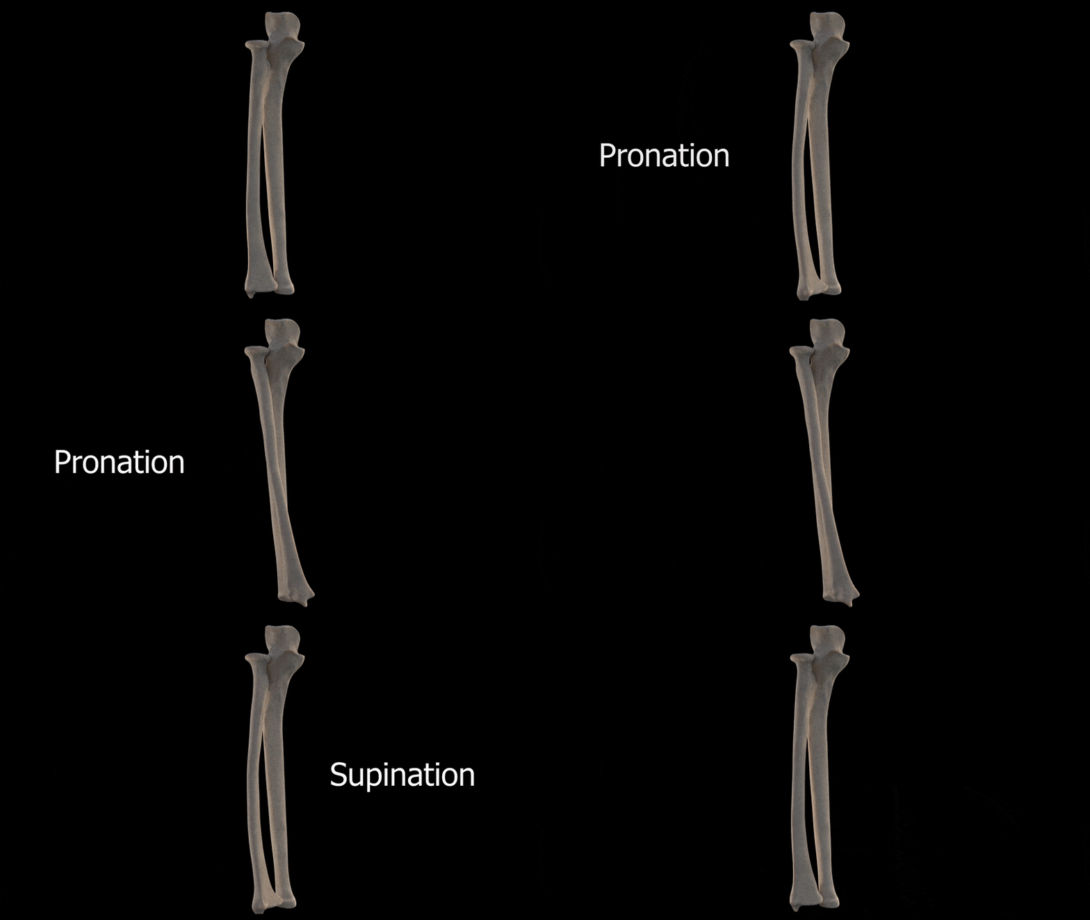
The first two muscles you will need to learn in this category are involved in two opposing functions of the forearm
- pronation
- supination
Use the names of each muscle to remember their functions.
Supinator
This is a short and deep muscle near the elbow in the posterior compartment of the forearm. It does not extend into the digits. Its antagonist is the pronator teres (see next muscle below).
Visit this AnatomyZone site. Press the 3D button. Scroll through the first two images using the arrows at top left. Press the up arrow on the right to open the 3D controls and use your mouse to rotate the forearm to view the origins and insertion of the supinator. Click on each bone for identification and review. See how it wraps around the radius and how this may help it carry out its function when it contracts.
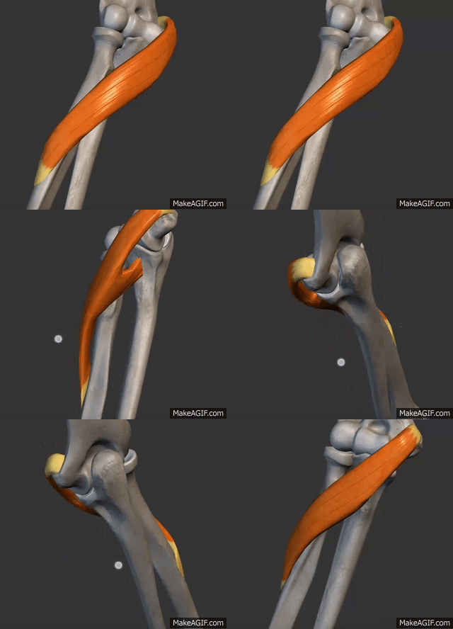
Pronator teres
The pronator teres muscle is one of four muscles within the superficial layer of the anterior compartment of the forearm. It does not extend into the digits. Try to see its sites of origin and insertion on the image to the right.
Visit this AnatomyZone site. Move the pronator teres around in 3D to observe it sites of origin and insertion. You can also click on the bones for identification. Move to page two of this site and identify the bones upon which the two heads of this muscle originate and insert. Based on this, are there any clues to what its function may be?
Muscles of the forearm that do extend into the digits
We will next consider two muscles that extend into the digits, as their names imply.
- flexor digitorum superficialis
- extensor digitorum
Flexor digitorum superficialis
This broad two-headed muscle occupies the intermediate layer of the anterior compartment of the forearm.
Visit this AnatomyZone site. Use your mouse to move the image around in 3D and take note of the anterior and posterior aspects using the thumb as your guide. You can also click on the bones for identification. Open the info panel on the left to view the labelled origin and insertion sites of this muscle. Make a note of this muscle’s sites of origin and insertion. Based on this, are there any clues to what its function may be?
Extensor digitorum
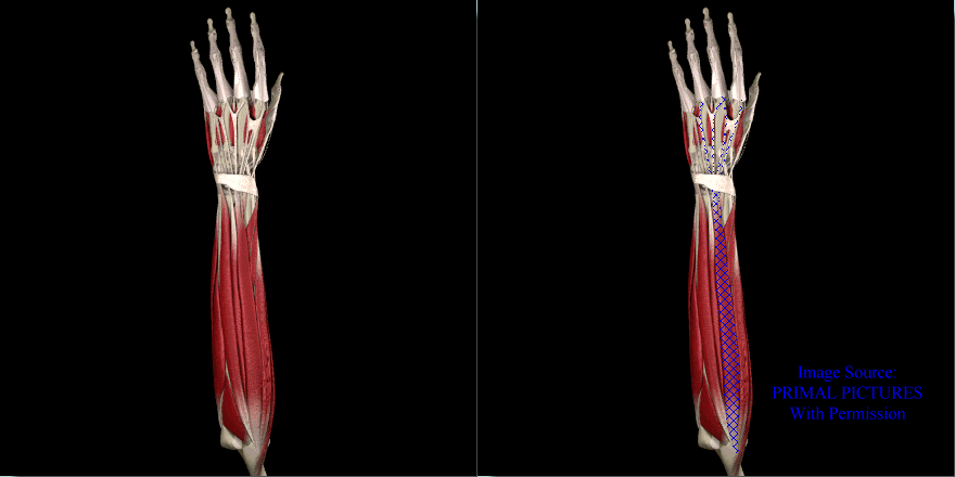
The extensor digitorum muscle is a superficial muscle of the posterior forearm present in humans and other animals.
Visit this AnatomyZone site. Move the image around in 3D to observe and make a note of this muscle’s sites of origin and insertion. You can also click on the bones for identification. Can you see whether the origin site is lateral or medial? Are there any clues to what its function may be?
