
Learning Objectives
By the end of this section, you will be able to:
Identify, locate, and describe the origin, insertion and function of the major upper and lower leg muscles.
Upper Leg Muscles
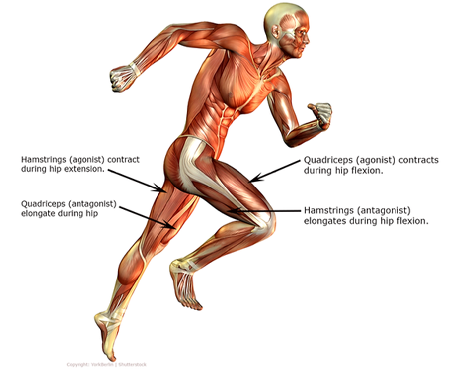
We will consider the following two muscle groups with opposing functions:
- quadriceps
- hamstrings
When the hamstring muscle group acts as the agonist (primary mover), the quadriceps serve as the antagonist (mainly counteracting the force generated by the agonist).
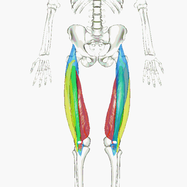
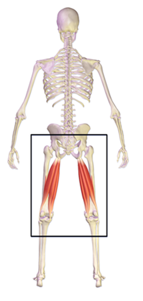
Like the forearm, the upper leg, or thigh, has a dense arrangement of many muscles. On the anterior side, the most prominent of the muscles are the sartorius muscle and the four muscles that make up quadriceps muscle group (the “quads”). You are responsible for learning about the quadriceps femoris group.
By BodyParts3D/Anatomography – BodyParts3D/Anatomography, CC BY-SA 2.1 jp, https://commons.wikimedia.org/w/index.php?curid=33808194
The quadriceps femoris name provides clues about these muscles. The latin root word ‘quad’ means four. The suffix ‘-ceps’ means head. Quadriceps literally means ‘four headed’. The four quadriceps muscles are arranged as follows:
- the rectus femoris is in the center,
- the vastus medialis on the medial side,
- the vastus lateralis on the lateral side and,
- the vastus intermedius is deep to the rectus femoris and hidden from view.
Together they cover the front and sides of the femur. Based on the description above, try identifying the muscles on the following H5P interactive.
wiki/File:Rectus_femoris_3D.gif
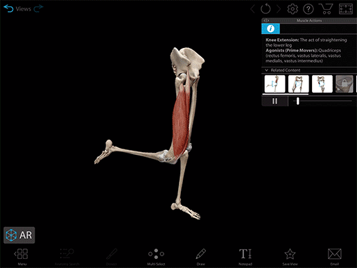
This muscle is part of the quadriceps group on the anterior compartment of the thigh. The two joints that it crosses provide clues to two of its functions.
Go to this AnatomyZone site to get a 3D image of the rectus femoris. Click on the 3D Model on the right of the screen. Make it full screen so you can see the origin and insertion sites of the muscle. Open the 3D controls by clicking the up arrow on the bottom right of the 3D model screen. Click on the second choice ‘change to rotate mode’ and use your mouse to rotate the image.
Watch this video on the rectus femoris, then test your recall of its origin, insertion and function using the separate toggles below the video.
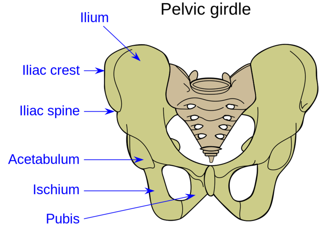
The origin of the rectus femoris is the iliac spine on the ilium of the pelvis.
The rectus femoris inserts as the quadriceps tendon on the upper border of the patella and continues as the patellar ligament to the tibia (tibial tuberosity).
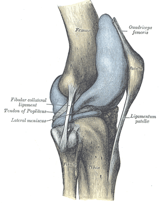
The rectus femoris functions in flexion of the hip joint and extension of the knee (–straightening the leg).
These three muscles form the remainder of the quadriceps femoris. As their names suggest, the vastus lateralis is on the lateral aspect of the thigh, the vastus medialis is medial, and the vastus intermedius is between the other two, deep to the rectus femoris.
Go to this AnatomyZone site. Locate the origin, insertion and function of the rectus femoris, vastus lateralis and medialis.
Watch this video on the vastus lateralis, then test your recall of its origin, insertion and function.
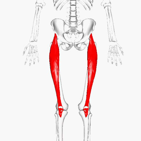
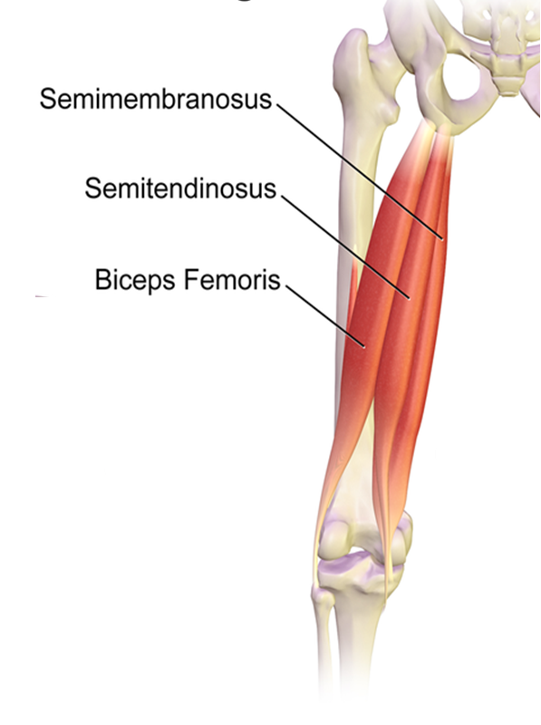
The following three long muscles on the back of the knee belong to the hamstring group:
- the semimembranosus is the most medial muscle,
- the semitendinosus is lateral to the semimembranosus muscle and,
- the biceps femoris has a long head and a short head and is the most lateral and deep of the hamstring muscles.
Based on the description above, try identifying the muscles in the following H5P exercise.
- Biceps Femoris
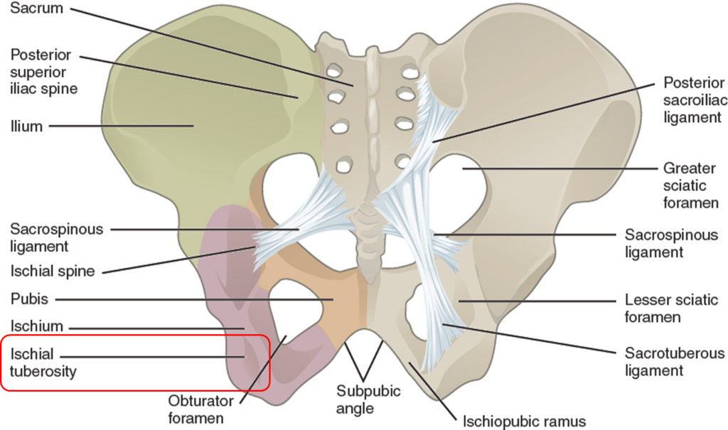
The semimembranosus and semitendinosus originate on the ischial tuberosity. The biceps femoris long head originates on the ishial tuberosity and the short head originates on the femur.
Insertion on the tibia:
- semimembranosus (posterior surface of medial tibial condyle)
- semitendinosus (proximal, medial surface of tibia)
- biceps femoris (lateral tibial condyle)
Insertion on the fibula:
- biceps femoris
https://en.wikipedia.org/wiki/Hamstring#/media/File:Pulled_Hamstring.png
Lower Leg Muscles
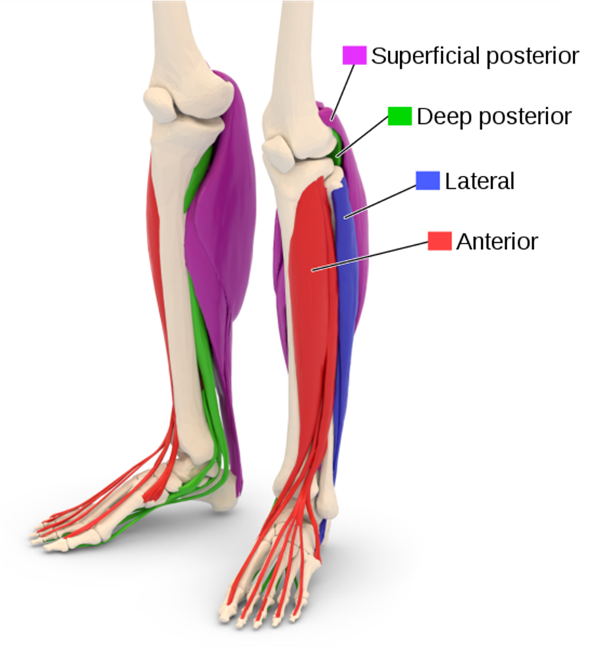
These muscles generally move the foot and toes. The leg has three compartments as shown in the image below: anterior, lateral and posterior (deep and superficial). We will consider one muscle of the anterior and two muscles of the posterior compartments.
Lower Leg-anterior compartment
The tibialis anterior is a long and thick muscle. It is the most medial muscle of the anterior compartment of the leg. Use the rotating image above to (1) first identify the origin, insertion and function of the muscle based on your previous knowledge of bone structure and (2) test your knowledge using the dropdowns below. (3) Lastly, open the ANATOMYZONE 3D modelling site below to reinforce your knowledge.
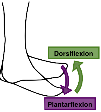
The tibialis anterior dorsiflexes the ankle and inverts the foot when it contracts. It opposes the gastrocnemius muscle.
ANATOMY ZONE 3D MODEL Visit this AnatomyZone site to rotate the muscle and observe its origin and insertion sites. Click on the + sign for the tibialis anterior and rotate the image to view the origin and insertion sites.
Lower Leg-posterior compartment
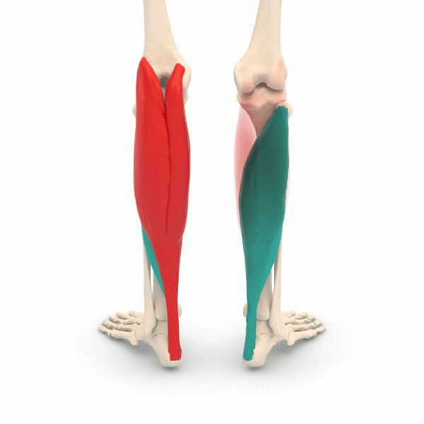
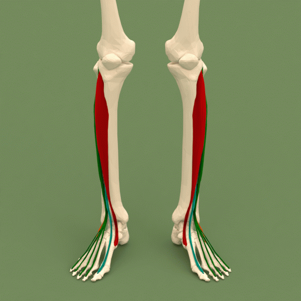
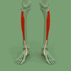
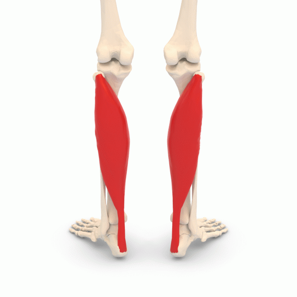
The most superficial and visible muscle of the calf is the gastrocnemius. This two-headed muscle runs from the back of the knee to the heel and is important in walking and posture.
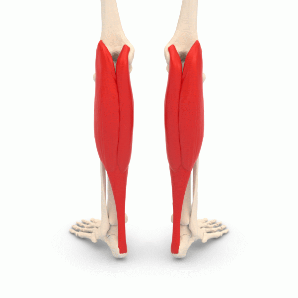
Tendons from both the gastrocnemius and the soleus merge to form a common tendon, the tendon of Achilles (calcaneal or gastrocnemius tendon) which inserts on the calcaneus.
The gastrocnemius flexes the lower leg at the knee joint and functions in plantar flexion of the foot (ankle extension).
Visit this AnatomyZone site to identify the origin and insertion of this muscle. Use the right/left arrows to scroll through each page. Rotate the image on page 2 to view the spatial relationship between the gastrocnemius and soleus muscles. Rotate the image on page 3 to view the origin and insertion sites of the soleus. Then, test your recall of its origin, insertion and function(s) below.
Test Your Knowledge
Try to correctly identify the lateral and medial heads of the gastrocnemius and two bones in the H5P exercises below.
Attributions
Gastrocnemius. Physiopedia contributors. Physiopedia (2022). Date retrieved 22 January 2024. https://www.physio-pedia.com/index.php?title=Gastrocnemius&oldid=298978
“BodyParts3D/Anatomography” by The Database Center for Life Science is licensed under CC BY-SA 2.1