Learning Objectives
By the end of this section, you will be able to:
- Identify key anatomical features of the radius and ulna, the two bones of the forearm
- Describe the structural differences and functional roles of each forearm bone
- Explain how the radius and ulna work together to support forearm movement, including flexion, extension, pronation, and supination
Introduction
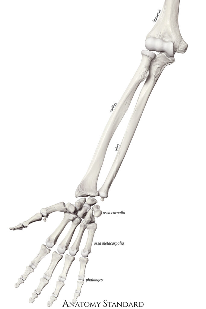
The forearm contains two long bones—the radius and the ulna—that work together to support movement and stability between the elbow and wrist. These bones are essential for both stability and mobility of the upper limb, enabling a wide range of movements.
- The radius is located on the lateral side (thumb side) of the forearm. Its disk-shaped head articulates with the capitulum of the humerus, allowing for the rotational movements of pronation and supination.
- The ulna, on the medial side (pinky side), forms the primary hinge joint at the elbow through its articulation with the trochlea of the humerus. It provides structural support and serves as a key attachment site for muscles involved in elbow and wrist motion.
Look at the figure. Determine whether the radius and ulna are lateral or medial. What trick will you use to remember this?
Use the interactive 3D models and labeled images in this section to explore the unique features of each bone. As you examine the radius and ulna, consider how their shapes and articulations contribute to the complex movements of the forearm.
The radius is a lateral bone of the forearm. It has a characteristic enlarged, disk-like head (a.k.a. radial head) that articulates with the capitulum of the humerus. This disk-like nature of the head makes possible the rotary pronation-supination of the hand.
Study Hint = Radius bone is on the same side as the thumb! Another fun hint = “Thumbs Up is Right!” = radius or hitching a ride with thumbs up.
Find the following landmarks on the radius shown in the image to the right:
Radial tuberosity: on the medial surface a few centimeters distal to the head; raised and rough surface that is the point of attachment of the biceps brachii muscle
Styloid process: enlarged distal process, articulates with the wrist. Is this process lateral or medial?
🌐 Explore in 3D:
Visit this AnatomyZone site to view the radius in 3D. Click the arrows in the info panel and scroll through all four views to identify each landmark. Move the bone around with your mouse. Using the thumb, as your landmark, make a note of whether each landmark is medial or lateral.


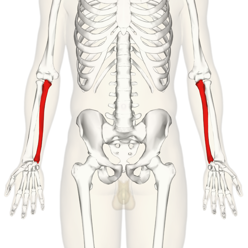
The ulna is the medial bone of the forearm, positioned on the side closest to the body (pinky side).
Proximal end
- The proximal end features the olecranon, which fits into the olecranon fossa of the humerus when the arm is extended. Just anterior to the olecranon is the trochlear notch, a large, C-shaped groove that articulates with the trochlea of the humerus. Together, these structures give the proximal ulna an appearance similar to an ice cream scoop, forming a strong hinge joint at the elbow joint.
Distal end
- Styloid process: Located on the posteromedial surface of the distal ulna, this projection can be easily felt through the skin at the wrist. Try locating this process on your own wrist for a tactile reference.
🌐 Explore in 3D:
Visit this AnatomyZone site to view the ulna in 3D. Click the arrows in the info panel and move the bone around with your mouse. Using the thumb, as your landmark, make a note of why the ulna is considered medial or lateral.


Elbow Joint
To the lateral side and slightly inferior to the trochlear notch is a small, smooth area called the radial notch of the ulna. This area is the site of articulation between the proximal radius and the ulna, forming the proximal radioulnar joint. The posterior and superior portions of the proximal ulna make up the olecranon process, which forms the bony tip of the elbow. the coronoid and olecranon processes (on the ulna) articulate into the coronoid and olecranon fossae (on the humerus), respectively.
Inspect the features of the elbow joint in the figure below.

Watch this Anatomy Zone video on the elbow joint.
Think about the shape of the olecranon and trochlear notch, then check your reasoning as you explore the 3D model & movie.