Bones of the Upper Arm

Learning Objectives

By the end of this section, you will be able to:

  • Identify the major anatomical landmarks of the humerus.
  • Describe the location and function of key features at the proximal, shaft, and distal regions.
  • Relate humerus structure to its articulations with the scapula, radius, and ulna.

The humerus

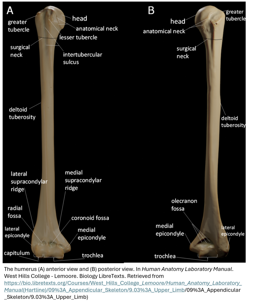
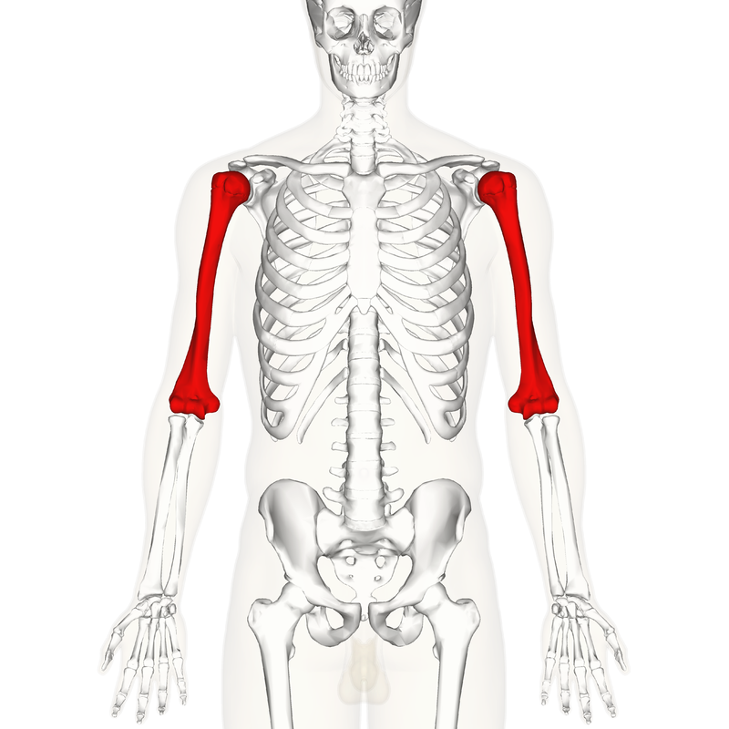
The humerus is the single bone of the upper arm region. This upper arm bone articulates proximally with the scapula and distally at the elbow with the radius and ulna. The proximal end is expanded into a head which articulates with the glenoid cavity of the scapula.

By Anatomography – en:Anatomography (setting page of this image), CC BY-SA 2.1 jp, https://commons.wikimedia.org/w/index.php?curid=23389521
Let’s first break down the key regions of the humerus.

Use the labeled images below to explore the major anatomical landmarks of the humerus. As you review each region—proximal, shaft, and distal—match the descriptions to the visual features shown.

Proximal End:

  • Head: Articulates with the glenoid cavity of the scapula.
  • Greater tubercle (lateral) and lesser tubercle (anterior): Prominent sites for muscle attachment.
  • Intertubercular sulcus (groove): Located between the tubercles; passageway for tendons.
  • Anatomical neck and surgical neck: Important clinical landmarks.

Shaft:

  • Deltoid tuberosity: Raised ridge on the lateral side; attachment site for the deltoid muscle.
  • Lateral and medial supracondylar ridges: Sites for muscle attachment.

Distal End:

  • Capitulum: Rounded lateral surface; articulates with the radius.
  • Trochlea: Medial surface; articulates with the ulna.
  • Radial fossa and coronoid fossa: Depressions for articulation during elbow flexion.
  • Olecranon fossa (posterior): Accommodates the olecranon of the ulna during elbow extension.
  • Medial epicondyle: Blunt projection felt on the inner elbow; the ulnar nerve passes behind it—pressure here causes the “funny bone” sensation.
  • Lateral epicondyle: Prominent process on the outer side of the distal humerus.

Check out this short video from AnatomyZone that walks you through the key landmarks of the humerus—from the head and tubercles to the epicondyles and fossae.

As you watch, try to match each feature to the labeled images above. Can you spot the same structures in the video that you’ll explore in 3D next?

Try this video below as well! (donated by BIOL 1594 student!). Open in full-screen mode for a better view.

Ready to test your observational skills? Use the 3D models below to identify the structures you’ve just learned about.

Activity 1: Explore the landmarks described above using Sketchfab’s humerus model.
Can you identify the various landmarks before clicking on the numbers?

Activity 2: Visit this AnatomyZone site to explore the humerus and its proximal and distal articulations in 3D. Observe the key landmarks described above while using the arrows to scroll through the info panel. Scroll through all seven pages.

Answer:
A fracture at the surgical neck can damage the axillary nerve, leading to weakness in shoulder abduction and loss of sensation over the deltoid region. This affects both mobility and muscle function.

Answer: One of the most important landmarks of the humerus is the head, because it forms the ball of the shoulder joint by articulating with the glenoid cavity of the scapula. This ball-and-socket joint allows for a wide range of motion in the upper limb, including rotation, flexion, extension, and abduction. Without the rounded head and its smooth articulation surface, shoulder mobility would be significantly limited.

Other key landmarks like the greater and lesser tubercles are also essential—they serve as attachment points for major shoulder muscles, contributing to strength and stability during movement.

Test Your Knowledge

Visit the Innerbody website (Upper Limb: Bony Landmarks) before trying the Beat the Clock exercises below. Click 3D rotate and zoom to begin your journey. Try identifying landmarks and bones before clicking on them —can you identify them based on what you’ve learned already?

Correct answer: Medial epicondyle of the humerus

  • Medial epicondyle — the large, prominent bump on the inner side of your elbow (easily felt). It serves as an attachment point for the forearm’s flexor muscles.
  • Lateral epicondyle — the smaller bump on the outer side of your elbow, where the extensor muscles of the forearm attach.

So when you touch your elbow, the medial epicondyle is the most prominent and palpable part of the humerus.

The “funny bone” isn’t actually a bone — it’s a nerve!

When you hit your funny bone, you’re actually bumping your ulnar nerve as it passes behind the medial epicondyle of the humerus (the bony bump on the inner side of your elbow).

That’s why:

  • You feel a tingling or electric shock sensation instead of a normal bruise, and
  • The tingling runs down your forearm into your ring and little fingers — the areas supplied by the ulnar nerve.

So, the “funny bone” = ulnar nerve at the medial epicondyle of the humerus.

Now it’s your turn! Use the interactive activities below to test your knowledge of landmarks of the humerus. How quickly can you label each part?

  1. Go to the purpose games site & press play. Use the figure provided in this section to identify various landmarks. See how fast you can get!
  2. Go to the purpose games site & press play. Use the figure provided in this section to label a few more landmarks. See how fast you can get!

Hartline, R. (n.d.). 9.3: Upper Limb. In Human Anatomy Laboratory Manual. West Hills College – Lemoore. Biology LibreTexts. Retrieved from https://bio.libretexts.org/Courses/West_Hills_College_-Lemoore/Human_Anatomy_Laboratory_Manual(Hartline)/09%3A_Appendicular_Skeleton/9.03%3A_Upper_Limb/09%3A_Appendicular_Skeleton/9.03%3A_Upper_Limb)