
Introduction
Whether you’re walking, running, or simply standing still, your lower limbs are doing a lot of work behind the scenes. From the powerful femur in your thigh to the intricate bones of your feet, this region is a marvel of structural engineering. In this section, you’ll explore the bones that support your body, enable movement, and absorb the impact of every step. Ready to take a closer look at the framework that keeps you moving?
Like the upper limb, the lower limb is divided into three regions. The thigh spans from the hip joint to the knee joint. The leg refers specifically to the area between the knee and the ankle. Distal to the ankle is the foot, a complex structure built for movement and support. Altogether, the lower limb contains 30 bones: the femur, patella, tibia, fibula, tarsal bones, metatarsals, and phalanges.
🌐 Explore in 3D
Before diving into the details below, take a moment to explore the lower limb bones on the Innerbody website. Click 3D rotate and zoom to begin your journey. Try clicking on different landmarks—can you identify them based on what you’ve learned already?
Learning Objectives
By the end of this section, students should be able to:
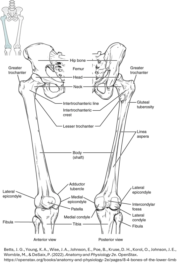
- Identify the femur and its key anatomical landmarks: head, neck, greater and lesser trochanters, and condyles.
- Understand the articulation of the femur with the pelvis at the acetabulum.
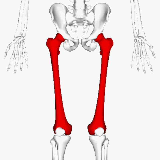
The femur isn’t just the longest bone in your body—it’s a powerhouse of support and movement. From its rounded head that fits snugly into the hip socket to the trochanters and condyles that anchor muscles and form joints, the femur is central to walking, running, and jumping. Let’s explore what makes this bone so strong and versatile.
1. 🌐 Explore in 3D
Visit this Sketchfab site to view a real human femur in 3D. Scanned and annotated by students in Dr. Eric Bauer’s human anatomy lab at Elon University, North Carolina, USA. You are responsible for recognizing the landmarks described below.
2. Femur Landmarks – Upper Leg (Thigh Bone)
- Head: Hemispherical structure at the proximal end; articulates with the acetabulum of the pelvis.
- Neck: Narrowed region just below the head; connects to the expanded proximal shaft.
- Greater Trochanter: Large, lateral projection for muscle attachment.
- Lesser Trochanter: Smaller, medial projection located slightly distal to the greater trochanter.
- Lateral and Medial Condyles: Prominent articular surfaces at the distal end of the femur; form part of the knee joint.
Test Your Knowledge
Learning Objectives
By the end of this section, students should be able to:
- Distinguish between the tibia and fibula in terms of size, location, and function.
- Recognize the patella as a sesamoid bone and describe its role in knee movement.
- Locate and describe the medial and lateral malleoli and their relationship to the ankle joint.
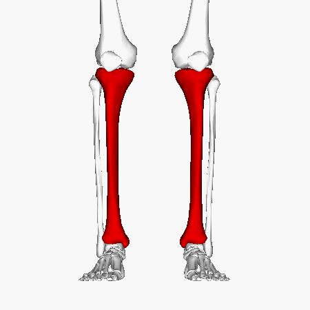
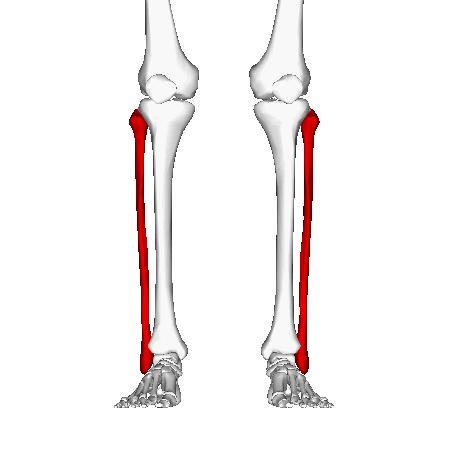
The tibia and fibula may look like a matched pair, but they play very different roles. One bears the weight, the other offers balance. Add the patella into the mix—a floating bone that boosts your knee’s efficiency—and you’ve got a dynamic trio that keeps you upright and moving. Time to meet the bones that carry you forward.
Start by reading the descriptions, then dive into the 3D models to learn about these two lower leg bones.
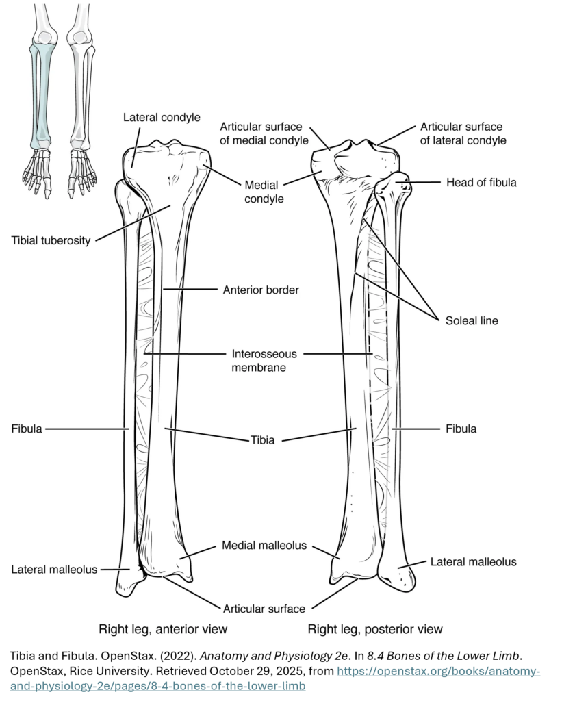
The tibia (shin bone) is the medial bone of the leg and is larger than the fibula, with which it is paired (see GIF). The tibia is the main weight-bearing bone of the lower leg and the second longest bone of the body, after the femur. The medial side of the tibia is located immediately under the skin, allowing it to be easily palpated down the entire length of the medial leg.
Landmarks
Proximal end: The two sides of this expanded area form the medial condyle and lateral condyle of the tibia. The top surface of each condyle is smooth and flattened, providing articular surfaces for the medial and lateral condyles of the femur—together forming the knee joint.
Distal end: The medial malleolus is the big bump you can feel on the inside of your ankle. It’s where the tibia flares out at the bottom. This smooth area, along with the bottom of the tibia, fits against the talus bone in your foot to help form the ankle joint.
🌐 Explore a real human tibia in 3D. Scanned and annotated by students in Dr. Eric Bauer’s human anatomy lab at Elon University, North Carolina, USA.
The fibula is the slender bone located on the lateral side of the leg (see GIF). The fibula does not bear weight. It serves primarily for muscle attachments and thus is largely surrounded by muscles. Only the proximal and distal ends of the fibula can be palpated.
Landmarks
Proximal end: The head of the fibula is a small, rounded bump at the top of the bone. It connects to the lower side of the lateral tibial condyle, forming the upper tibiofibular joint.
Distal end: The lateral malleolus is the bony bump you can feel on the outside of your ankle. It’s part of the lower end of the fibula. The smooth inner surface of the lateral malleolus connects with the talus bone in the foot to help form the ankle joint. The bottom of the fibula also fits into a notch on the tibia.
🌐 Explore a real human fibula in 3D. Scanned and annotated by students in Dr. Eric Bauer’s human anatomy lab at Elon University, North Carolina, USA.
Use the landmarks you explored above to see how the tibia and fibula fit together.
Whether you prefer interactive 3D models or static figures, both are available here to help you visualize the articulation of these two lower leg bones.
Learning Objectives
By the end of this section, students should be able to:
- Identify the talus and its articulation with the tibia and fibula.
- Understand the structural role of the foot bones in weight-bearing and movement.
Your foot is a complex structure built for both stability and flexibility. With bones that form arches, absorb shock, and adapt to uneven terrain, it’s a biomechanical marvel. From the ankle’s talus to the tips of your toes, each bone plays a part in keeping you grounded—literally.
Regions of the foot
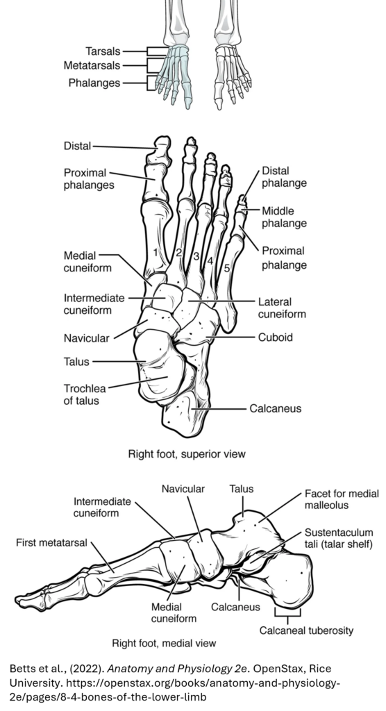
Tarsals (ankle)
- Made up of seven bones.
- Includes the calcaneus (heel bone), which sticks out at the back for muscle attachment.
- The talus sits on top and connects with the tibia and fibula to form the ankle joint.
- (You don’t need to memorize the names of the other tarsal bones.)
Metatarsals (instep)
- Like the hand’s metacarpals, there are five long bones in the foot.
Phalanges (toes)
- Similar to fingers: 14 bones total.
- Each toe has three bones, except the big toe, which has two.