
Learning Objectives
By the end of this section, students should be able to:
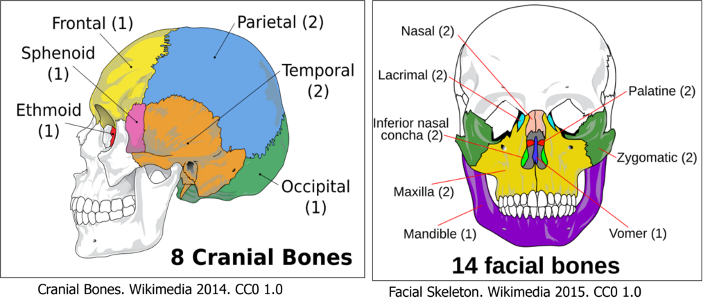
- Identify and describe the 8 cranial and 14 facial bones of the skull.
- Locate and name key foramina, processes, and sutures on the skull.
- Recognize the location and purpose of four paranasal sinuses.
- Distinguish between adult and fetal skull features, including fontanelles.
Cranial and Facial Bones
The skull consists of twenty-two bones that are divided into two divisions. These bones have complex shapes, and they fit together like the pieces of a very intricate jigsaw puzzle. You’ll begin by examining the skull as a whole from multiple viewpoints, then explore specific features of each bone.
To support different learning styles, each section on this page combines labeled figures, descriptive text, rotating GIFs, 3D models, and interactive H5P activities—giving you multiple ways to engage with the material and build your understanding & recall skills. Let’s get started!
Use these resources to preview the skull’s anatomy before exploring individual bones and interactive tools. You’ll be expected to identify key bones, sutures, and openings. These labeled illustrations from Betts et al. (2022) provide detailed views of the skull, including cranial and facial bones, sutures, and foramina.
💡 Use these images to reinforce your understanding of bone names, connections, and key anatomical landmarks.
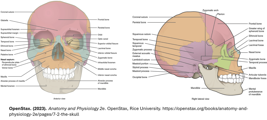
📎 Click here for a detailed description of the human skull (Betts et al., 2022). This OpenStax A&P chapter provides in-depth descriptions of each bone and its features.
📎Click here for another detailed description of the human skull (Lange et al., 2025)
Both OpenStax and LibreTexts chapters provide in-depth descriptions of each bone and its features with slightly different visuals.
💡 Tip: These resources are great for reviewing before or after using the dropdowns and 3D models below.
🦴 Learn the Skull Bones
Explore the 8 cranial bones and 14 facial bones summarized in the figure one by one using the dropdowns below. Each dropdown includes a summary of the bone’s location, features, and functions, along with rotating skulls and links to interactive 3D tools to help you visualize its structure, connections and relative position.
💡 Tip: You can also scroll down to the Summary of Interactive 3D Skull Models section —each with a QR code so you can easily open them on your phone while reviewing content on your laptop. Then, reinforce your learning with the Test Your Knowledge activities.
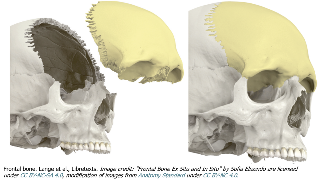
Location: Anterior and superior portion of the cranium. A single bone that forms the forehead.
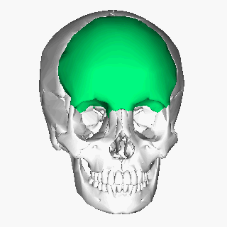
Description:
- Begins as two bones that typically fuse around age 8.
- Forms the eyebrow ridges and the ridge above the nose.
- Extends inferiorly into the orbit of the eye (eye socket).
- Contains the supraorbital foramen.
💡 Tip: Go to Interactive 3D Tools to view the frontal bone, its relative location and various articulations. Test your identification skills before heading to the Test Your Knowledge section.
💡 Interactive 3D Tip:
- Visit the InnerBody Skull Explorer and activate the 3D rotate & zoom tool. Highlight the frontal bone in blue. While highlighted, rotate skull to view how the frontal bone articulates with surrounding bones!
- Visit AnatomyTOOL Exploded Skull! Click on the frontal bone to confirm your identification. Take note of its relative location in the cranium.
- Use Friso Jansen’s 3D model to test your identification of the frontal bone.
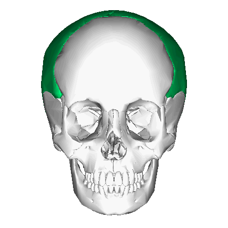
Location: Posterior to the frontal bone; form the superior and lateral surfaces of the cranium.
Description:
- Meet along the midline to form the sagittal suture.
- Each parietal bone articulates anteriorly with the frontal bone, inferiorly with the temporal bone and posteriorly with the occipital bone.
Challenge Yourself:
Can you describe the anatomical position of the parietal bones using directional terms in relation to both the frontal and occipital bones?
💡 Hint: Think superior, posterior, and lateral!
💡 Tip: Go to Interactive 3D Tools to view the parietal bones, their relative locations and their various articulations. Test your identification skills before heading to the Test Your Knowledge section.
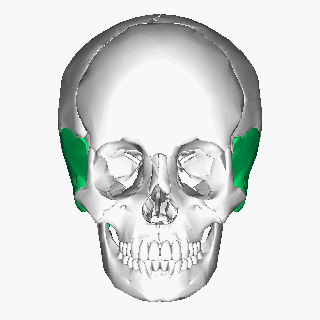
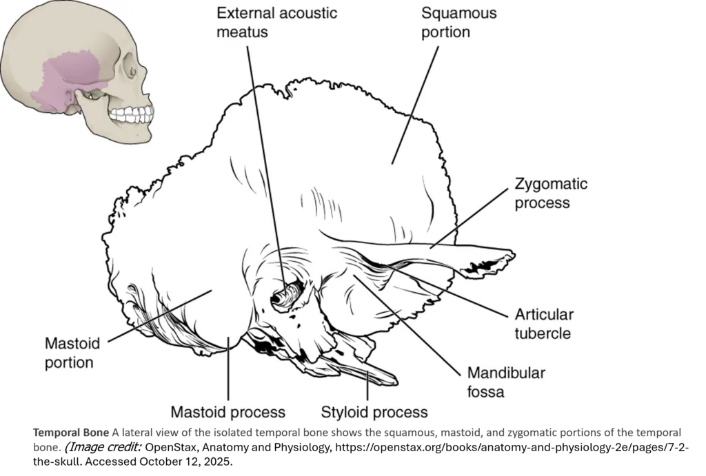
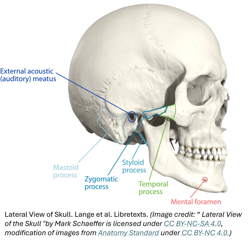
Location: Lower lateral walls on both sides of the cranium.
Description:
- Articulate with the mandible (lower jaw) via the mandibular fossa:
- a deep, oval-shaped depression located on the external base of the skull, just in front of the external acoustic meatus.
- Mandibular condyle is the rounded portion of mandible that articulates in the fossa to make the temporomandibular joint allowing for mouth movement.
- Posterior to the fossa is the external auditory meatus (ear canal).
- Temporal bones have three processes for muscle attachment:
- Zygomatic process
- projects anteriorly to form posterior portion of zygomatic arch
- Styloid process
- an elongated, downward bony projection on external base of the skull, so named because of its resemblance to a stylus (a pen or writing tool)
- Mastoid process
- projects inferiorly and can easily be felt on the side of the head just behind your earlobe. Serves as an attachment site for neck muscles including the sternocleidomastoid, which you’ll learn about later.
- Zygomatic process
💡 Tip: Use interactive tools to view the temporal bone as a whole, its features described above and its articulation with the mandible.
- First, launch Friso Jansen’s Labeled Skull (Sketchfab) and identify the temporal bone. Make note of its relation to surrounding bones.
- Then, launch BioDigital’s female skull. Highlight the temporal bone & then click on the features described above.
Test your identification skills before heading to the Test Your Knowledge section.
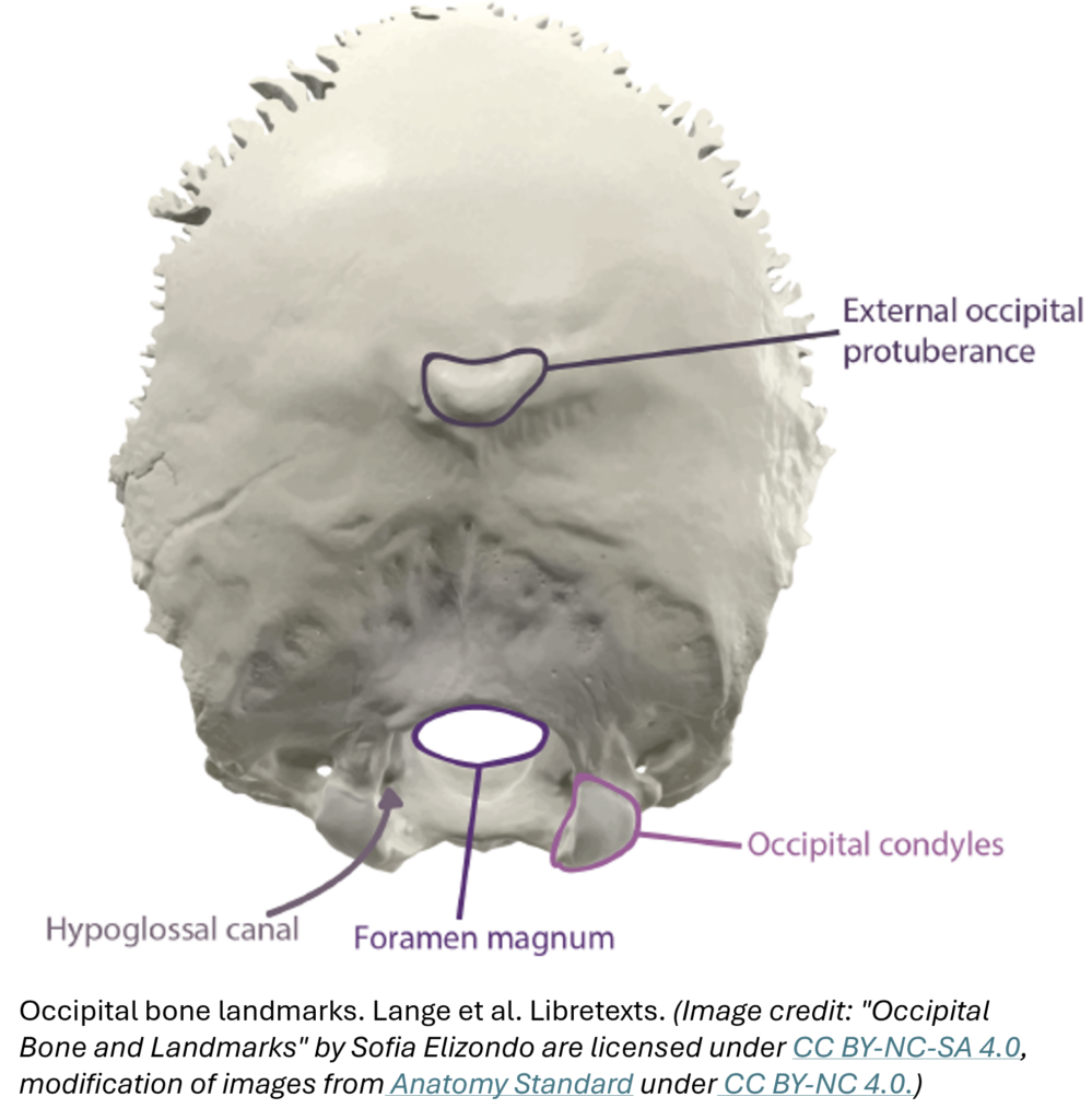
Location: Posterior and basal or inferior part of the cranium.
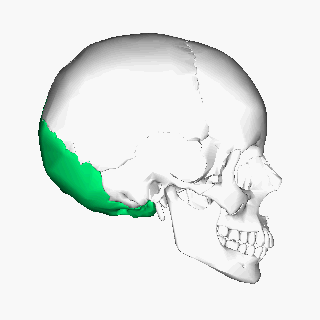
Description:
- Contains the large opening of the foramen magnum allowing passage of the spinal cord
- Features paired occipital condyles on either side of the foramen magnum
- Articulates with the atlas (C1 vertebra) to support and allow nodding motion of the skull.
💡 Interactive 3D Tip: Take note of directional positioning & bone relationships as you explore.
- Use BioDigital’s female skull model to highlight the temporal bone. Then click on the features described in the figures and text above.
- Use Eric Bauer’s 3D occipital bone model (sketchfab) to explore the bone’s key features in isolation.
Location: Anterior to the temporal bone; inferior to frontal and parietal bones.
Description:
- Acts as a keystone bone, connecting multiple cranial and facial bones.
- Forms the posterior wall of the eye orbit.
- Resembles a butterfly wing when viewed from inside the cranium.
- Contains sphenoidal sinuses that drain into the nasal cavity.

💡 Interactive 3D Tip:
- Visit the InnerBody Skull Explorer and use the selection tool to highlight the sphenoid bone in blue. While highlighted, rotate skull to view the butterfly-shaped wings and observe how the sphenoid articulates with surrounding bones!
- Visit AnatomyTOOL Exploded Skull! Click on the sphenoid bone to confirm your identification. Take note of its central location.
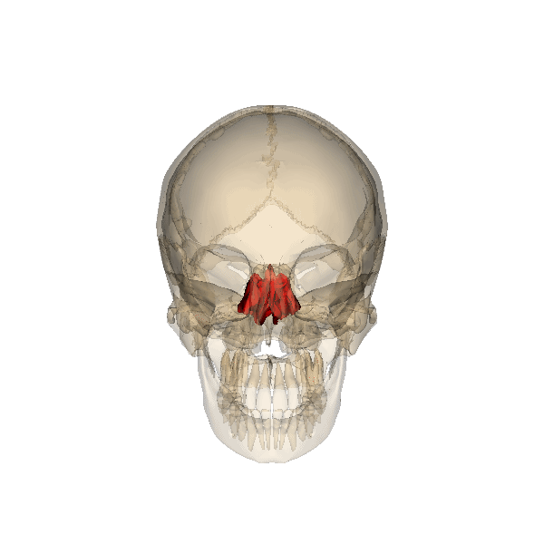
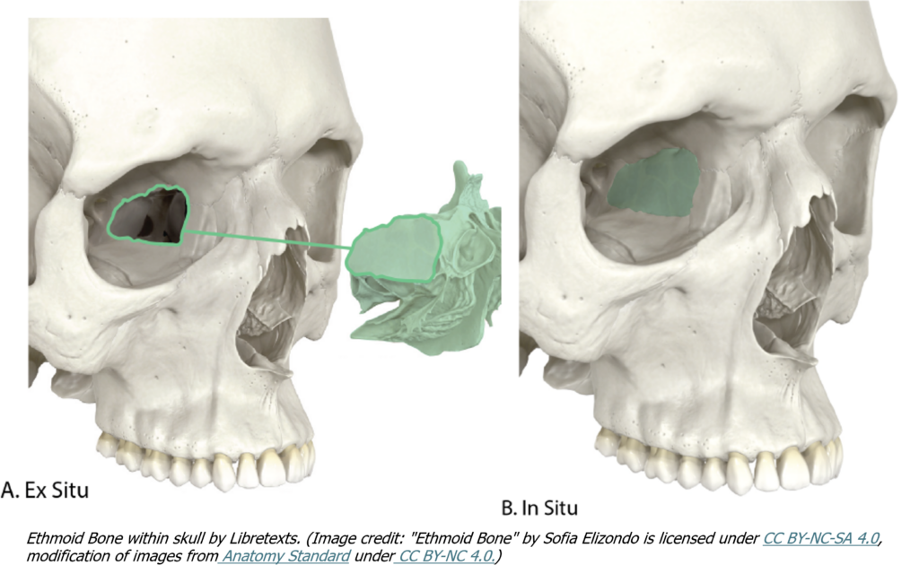
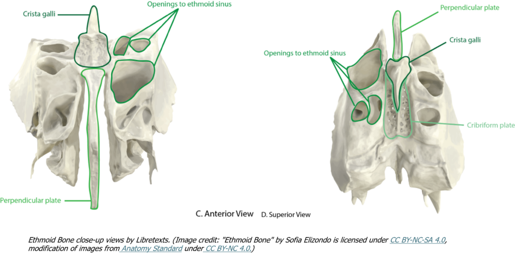
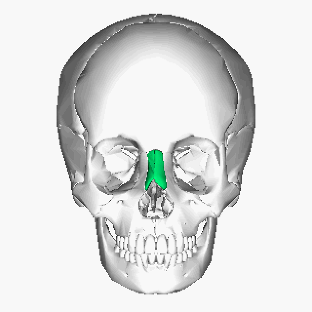
Location: Between the eye orbits; anterior cranial floor.
Description:
- Contains sinuses and forms part of the nasal septum.
- Has two bony shelves called superior and middle nasal conchae.
💡 Interactive 3D Tip:
- Visit Friso Jansen’s 3D skull model and locate the ethmoid bone. Click on the numbered label to confirm your identification, and take note of its central position within the skull and its relationship to surrounding bones.
- Visit BioDigital’s female skull. Highlight the ethmoid bone & then click on the information icon to read about it. What is it position in relation to the frontal bone?
- Visit AnatomyTOOL Exploded Skull! Click on the ethmoid bone to confirm your identification. Take note of its central location anterior to the sphenoid.
- Visit InnerBody. Activate the 3D rotate tool and try to identify the sphenoid and the ethmoid bones to see their relative positions.
Location: Bridge of the nose.
Description:
- Fused small rectangular/oblong bones forming the bridge of the nose.
- Cartilage below forms most of the nose.

💡 Interactive 3D Tip:
1. Visit the InnerBody Skull Explorer and click on the nasal bones to highlight them in blue. Rotate the skull to take note of their position in relation to other facial bones.
2. Visit InnerBody Nasal Bones to read a description of these bones.
3. Visit AnatomyTOOL Exploded Skull! Try to find the nasal bones! Note their size relative to other skull bones.
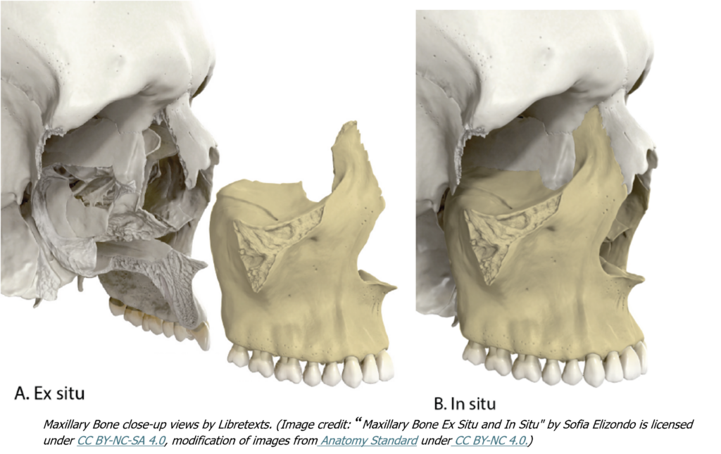
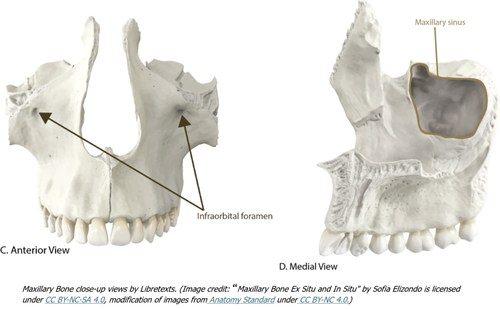
Location: Upper jaw.
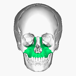
Description:
- Form the upper jaw, hard palate, floor of eye orbit, and nasal cavity walls.
- Support upper teeth in alveoli (socket).
- Contain infraorbital foramina (supplying nerves & blood vessels to the nose).
💡 Interactive 3D Tip:
1. Visit Whole Skull Anatomy – 3D Atlas of Neurological Surgery and identify the numbered maxillary bone.
2. Visit InnerBody’s Human Skull. Activate the 3D rotate tool and click on the maxillae bones to highlight them in blue. Rotate the skull to view its relative positions and various articulation points.
3. Visit AnatomyTOOL Exploded Skull! Identify the maxilla & rotate the skull to see its relationship with the jaw & teeth.
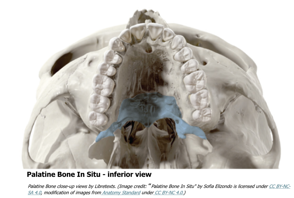
Location: Posterior third of the hard palate (hard to see).
Description:
- L-shaped bones forming part of the nasal cavity and eye orbit floor.
- The plates from the right and left palatine bones join together at the midline to form the posterior quarter of the hard palate.
The palatine bones are best seen in an inferior view of the skull and hard palate.

💡 Interactive 3D Tip:
1. Visit Whole Skull Anatomy – 3D Atlas of Neurological Surgery. Click around to get the skull to rotate to the inferior side to identify the palatine bone.
2. Visit AnatomyTOOL Exploded Skull! Rotate the skull to its inferior side. Identify the palatine bone & note its position in relation to other facial and cranial bones.
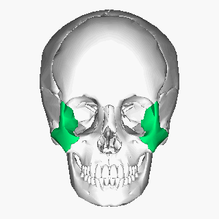
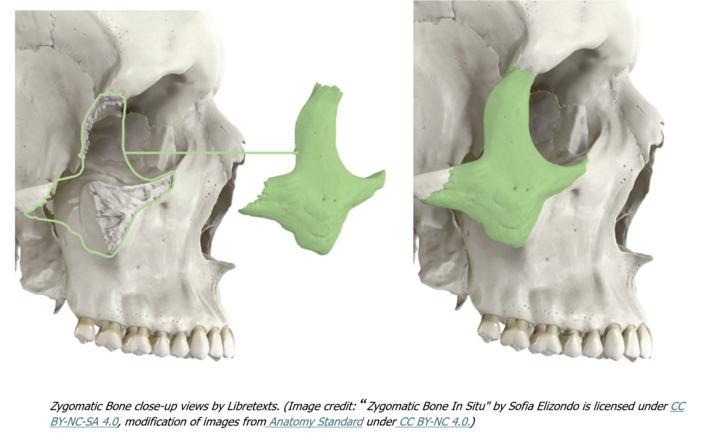
Location: Cheek and lateral/inferior eye orbit.
Description:
- Form the cheek prominence and eye orbit surface.
- Temporal process projects posteriorly to form a short extension that forms the anterior portion of the zygomatic arch
💡 Interactive 3D Tip:
1. Visit Friso Jansen’s Labeled Skull (Sketchfab) to identify the zygomatic bone. View the relationship between the zygomatic & temporal bones.
2. Visit Whole Skull Anatomy – 3D Atlas of Neurological Surgery. Click around to identify the zygomatic bone. Take note of how its temporal process articulates with the temporal bone to form the zygomatic arch.
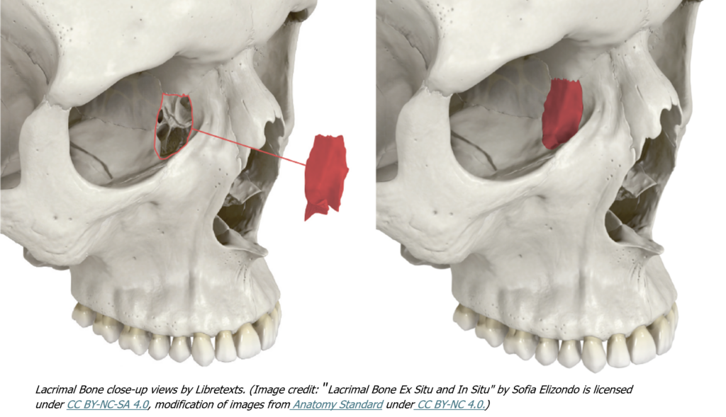
Location: Medial eye orbit.
Description:
- Small rectangular bones between maxilla and anterior portion of the ethmoid.
- Tears keep the eye’s surface moist and drain through the nasolacrimal canal at its inner corner.
💡 Interactive 3D Tip:
1. Visit the InnerBody Skull Explorer and click on the lacrimal bone to highlight it in blue. Rotate the skull to take note of its position in relation to other facial bones.
2. Visit AnatomyTOOL Exploded Skull! Try to find the lacrimal bone!
3. Now visit Whole Skull Anatomy – 3D Atlas of Neurological Surgery for a more authentic view of the lacrimal bone.
Location: Lateral nasal cavity.
Description:
- Thin, curved bones that divide the nasal cavity into air channels.
- Covered with membranes.
- Divide the nasal cavity into the channels through which air flows to be warmed and moistened.

💡 Interactive 3D Tip:
1. Visit the InnerBody Skull Explorer. Use the 3D rotation tools to hunt for the inferior nasal conchae in this model.
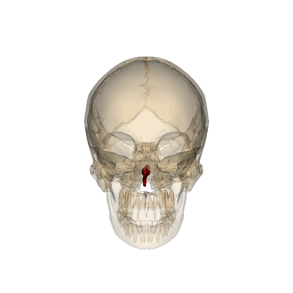
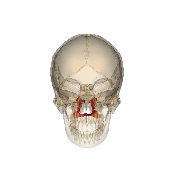
Location: Posterior median line of nasal cavity.
Description:
- Best seen when looking from behind into the posterior openings of the nasal cavity
- Joins maxillae, palatines, ethmoid, and sphenoid.

💡 Interactive 3D Tip:
1. Visit Friso Jansen’s Labeled Skull (Sketchfab) to see how the vomer divides the nasal cavity. Vomer isn’t labelled-can you find it?
2. Visit Whole Skull Anatomy – 3D Atlas of Neurological Surgery for a more authentic view of the vomer. Note its relation to the palatine bone.
3. Visit the InnerBody Skull Explorer. Activate the 3D rotate tool & find the vomer to highlight it in blue. Rotate the skull to take note of its position in relation to other facial bones.
4. Visit AnatomyTOOL Exploded Skull! Find the thin vomer bone!
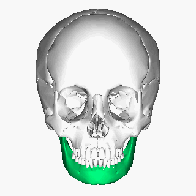
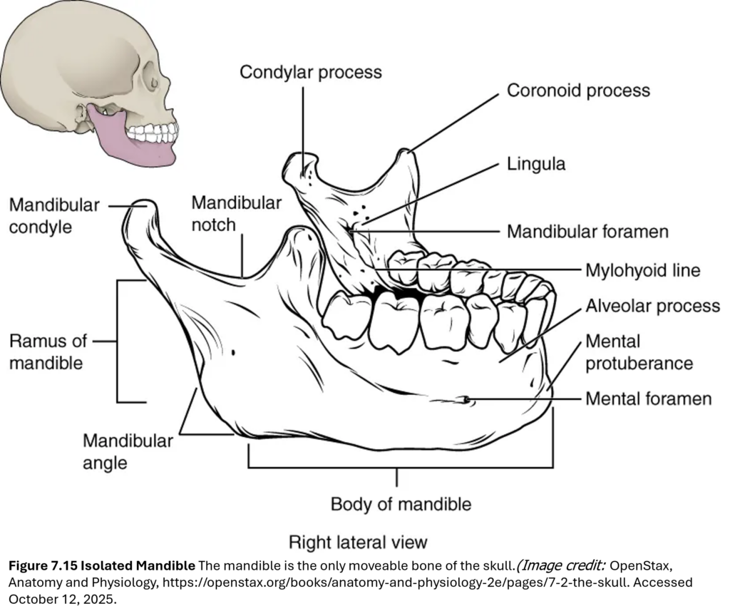
Location: Lower jaw.
Description:
- Only movable skull bone.
- Contains body and rami (ramus – singular; rami – plural):
- Each ramus is a vertically oriented branch consisting of the following:
- Mandibular condyle—articulates in mandibular fossa of temporal bone to form the temporomandibular joint
- Coronoid process for muscle attachment
- Mandibular notch
- Teeth sit in alveoli.
- Mandibular foramina present on medial surfaces of the rami.
- Two mental foramina on the outer lateral surfaces of the body that provide openings for a sensory nerve that supplies the chin.
- Mental protuberance projects forward from the inferior margin of the anterior mandible that forms the chin (mental=”chin”).

💡 Interactive 3D Tip:
1. Visit Friso Jansen’s Labeled Skull (Sketchfab). Identify the mandible & see how it articulates with the temporal bone.
2. Visit the InnerBody Skull Explorer. Activate the 3D rotate tool & click on the mandible to highlight it in blue. Rotate the skull to take note of its position in relation to other facial bones. What other features do you see in the 3D model that are described above?
Use these interactive models to rotate, zoom, and examine the skull from multiple angles. These tools help you visualize how cranial and facial bones fit together and where key sutures and foramina are located.
💡 Tip: Open these tools in a separate browser tab or on your phone so you can explore while reviewing bone descriptions provided above.
📌 What to look for while exploring:
- Cranial bones
- Facial bones
- Major sutures
- Foramina

Click the selection tool arrow and choose “Hide” to remove the colored muscles and focus on the bones.
Rotate the skull and click on individual bones to view their names and descriptions.
💡 Great for reviewing cranial vs. facial bones and exploring bone relationships interactively.

View the human skull as it separates into individual, color-coded bones—each clearly labeled for easy identification.
💡 Perfect for studying how bones connect, identifying sutures, and visualizing the spatial relationships between cranial and facial bones.

👉 Friso Jansen’s Labeled Skull (Sketchfab)
Explore a clean, anatomically accurate 3D model of the human skull with clearly labeled cranial and some facial bones. Rotate and zoom to examine bone shapes, connections, and spatial relationships from multiple angles.
💡 Excellent for learning bone names and practicing identification before testing your knowledge.

👉 Whole Skull Anatomy – 3D Atlas of Neurological Surgery
Explore a highly detailed 3D model of the human skull featuring labeled bones, sutures, and foramina—including the supraorbital foramen.
💡 Ideal for deep dives into skull anatomy, though it includes more structures than required for this course—focus on the bones bolded in your lab manual.
👉Bony Landmarks – BioDigital Human Interactive Model

Make a free account. Explore a full-body 3D model of the female and male adult skeletons and skulls with labeled bony landmarks, including key projections, depressions, and openings.
💡 Ideal for reviewing bone markings in context. Focus on the bolded features in your lab manual, and use the zoom and rotate tools to examine how these landmarks relate to muscle attachments, joints, and clinical assessments.
🔎 Note: The supraorbital foramina may not be visible in all digital modes.
✅ Test Your Knowledge
Ready to test your skills? Use these activities to check your ability to identify skull bones before moving on to the paranasal sinuses.
You’ll be expected to identify key bones, sutures, and openings. Use these models to test your ability to locate and name a variety of cranial and facial bones, as well as major foramina and sutures. Quickly review and quiz yourself on labeled bones and key features. Great for self-checks before & after lab!

👉 Friso Jansen’s Labeled Skull (Sketchfab)

How fast can you identify skull bones?
👉 Start the game. Good luck!
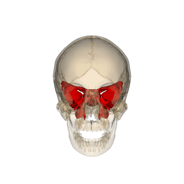
Paranasal Sinuses
The paranasal sinuses are hollow, air-filled spaces located within specific bones of the skull (Lange et al., 2025; Betts et al., 2022). Each sinus is named for the bone it occupies—paranasal literally means “next to the nasal cavity.”
These sinuses:
- Communicate with the nasal cavity
- Are lined with nasal mucosa
- Help lighten the skull by reducing bone mass
- Add resonance to the voice
They’re tucked inside bones you might not expect—like the sphenoid and ethmoid. Ready to explore where they’re hiding?
💬 Challenge:
See which sinus surprises you the most? Can you explain why it’s located there or how it might affect your voice or breathing?
- Frontal sinus: Most anterior, located above the eyebrows in the frontal bone.
- Maxillary sinuses: Largest, found in the maxillary bones below the orbits.
- Sphenoid sinus: Most posterior, deep within the body of the sphenoid bone.
- Ethmoid air cells: Multiple small spaces between the medial wall of the orbit and the lateral wall of the upper nasal cavity (Betts et al., 2022).
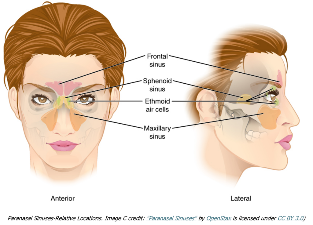
The paranasal sinuses are named for the skull bone that each occupies.

🧠 Quiz Yourself!
Can you identify all four paranasal sinuses in this 3D model by UMich Sketchfab ?
Try this:
- Locate the frontal, ethmoid, sphenoid, and maxillary sinuses.
- Which sinus is most anterior?
- Which one lies deepest within the skull?
- Rotate the model to view the sinuses from different angles.
💡 Tip: Use the labels and transparency features in the UMich Sketchfab model to help visualize their positions and connections.
✅ Test Your Knowledge
Ready to test your skills? Use these activities to check your ability to identify the paranasal sinuses before moving on to the fetal skull.
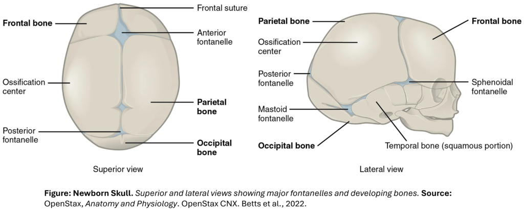
Fetal Skull
At birth, the skull is not fully ossified. The fontanelles—flexible regions of fibrous connective tissue—allow for distortion during birth and accommodate rapid brain growth.
Key Fontanelles to Identify:
- Anterior fontanelle: Diamond-shaped, between frontal and parietal bones.
- Occipital fontanelle: Between parietal and occipital bones.
- Sphenoidal & mastoid fontanelles: Seen on each inferior side.
Fontanelles close at various ages, but ossification completes by age two.
🌐 Interactive Tools -Explore the fetal skull in 3D
Now that you’ve reviewed the key fontanelles, take a closer look at a real fetal skull using this high-resolution 3D model.
👶 Featured Model: Human Fetal Skull by Eric Bauer
- Model : Real fetal skull captured via photogrammetry (Agisoft Photoscan)
- Features: High-resolution, rotatable in 3D, based on an actual specimen
- Use Case: Excellent for anatomical realism and studying fontanelles and sutures
🔗 View on Sketchfab

💬 Fetal Skull Challenges:
- Spot the Fontanelles:
Can you identify the four major fontanelles on the fetal skull? What do you notice about their shape and location? - Shape & Location:
Do the fontanelles look the same? How do their shapes and positions differ? Try describing one in your own words. - Pick a Favorite Fontanelle:
Which fontanelle stands out to you the most? Is it the size, the location, or what it allows the skull to do?
Ready to apply what you’ve learned? Use the tools in Test Your Knowledge below to label the fontanelles and test your speed!
✅ Test Your Knowledge
Before you wrap up, test your knowledge with the labeling challenge and timed activity!