A. Divisions of the Human Skeleton

Learning Objective

Differentiate between the axial and appendicular skeletons and identify their major components using diagrams, skeletal models, and digital tools.

Skeletal Divisions

The human skeleton protects important body regions and anchor muscles, making coordinated movements possible.  The usual 206 bones are grouped into the following two major divisions

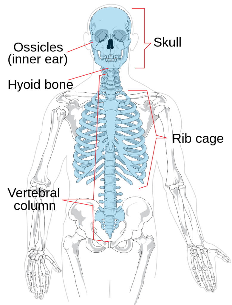
  1. The axial skeleton consists of the bones of the skull, vertebral column and thoracic cage (Figure 1a). You’ll begin by learning these foundational structures.
Figure 1a. The axial skeleton. (image by LadyofHats Mariana Ruiz Villarreal; https://en.wikipedia.org/wiki/Axial_skeleton)

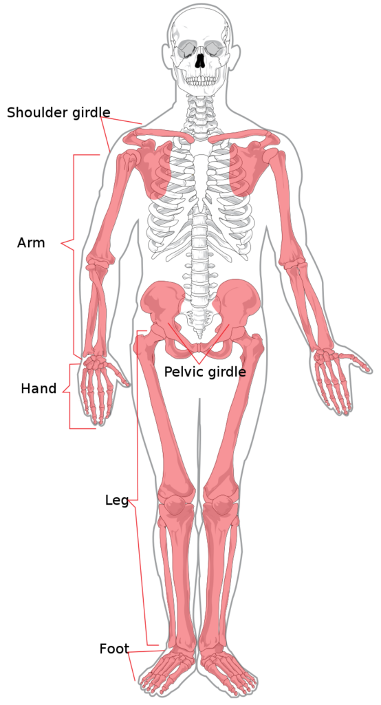
2. The appendicular skeleton is comprised of the bones of the arms and legs, and the two pelvic and shoulder girdles which anchor the limbs to the axial skeleton (Figure 1b).

Figure 1b. The appendicular skeleton (image by LadyofHats Mariana Ruiz Villarreal; https://en.wikipedia.org/wiki/Appendicular_skeleton)

Use the tools below to explore full-body 3D models of the human skeleton. These resources help you visualize how the axial and appendicular skeletons connect and function together.

👉 Explore the full-body 3D skeleton (MSD Manuals)

👉 Learn more about skeletal anatomy (InnerBody)

📌 What to do:
• Activate the 3D tools in both sites.

• Rotate and zoom in on the entire skeleton
• Try to identify axial vs. appendicular bones in real time—click on a bone for its name.
• Use this tool throughout the course for review and orientation

🔍 Challenge Yourself:

Use the interactive 3D skeleton above to identify and label five bones from the axial skeleton and five bones from the appendicular skeleton. Can you spot the differences in their location and function?

Ready to flex your anatomy muscles? Let’s see how well you can distinguish between the axial and appendicular skeletons!

Let’s put your visual skills to the test!

In the activities below, you’ll be shown full-body skeletal images. Your task is to decide which image represents the axial skeleton and which represents the appendicular skeleton. Use what you’ve learned from diagrams, models, and the 3D skeleton to guide your choices.

Activity 1: Look closely at the skeleton. Does it highlight the central framework—skull, spine, and rib cage or the limbs and girdle?

Activity 2: Examine these two skeletons. Are the limbs and girdles the focus? Are the skull, spine, and rib cage the focus?

The axial skeleton forms the central framework of the body—protecting vital organs like the brain, heart, and lungs, and providing a strong, stable axis for posture and support. It helps us stay upright and shield critical systems.

The appendicular skeleton is all about movement. It includes the limbs and the girdles that attach them to the axial skeleton, allowing us to walk, reach, grasp, and interact with our environment. This division makes it easier to study how bones work together to support both protection and mobility.