Learning Objectives
By the end of this section, you will be able to:
- Identify the anatomical features of a long bone.
- Describe the general structure and function of long bones.
- Explain the roles of compact bone, spongy bone, and bone membranes.
Introduction
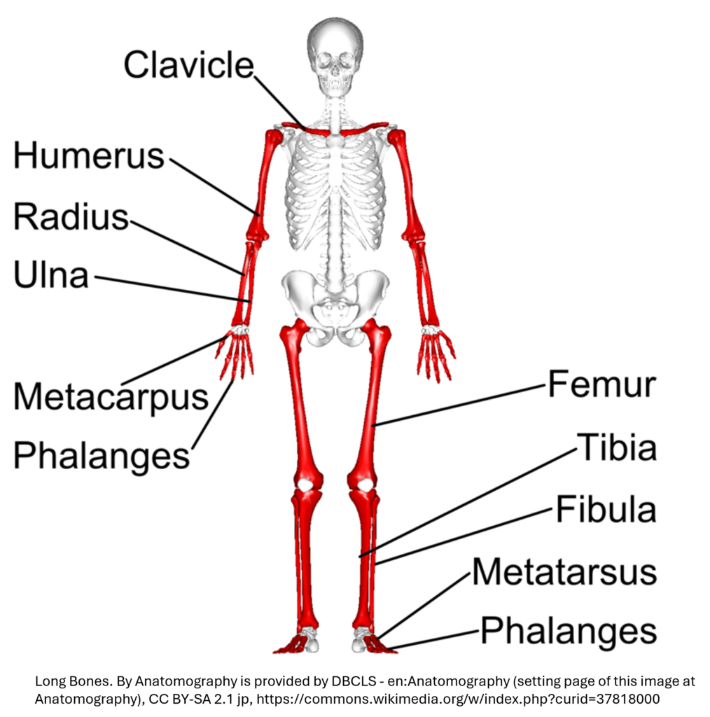
Long bones are those that are longer than they are wide, and they include bones in the arms, legs, fingers, and toes. These bones play a vital role in movement and support, especially the femur and tibia, which bear most of the body’s weight during daily activities.
In this section, you’ll explore the gross anatomy of a typical long bone, focusing on key structural regions like the diaphysis (shaft), epiphyses (ends), and surrounding layers such as the periosteum.
Understanding these features is essential not only for grasping how bones grow, repair, and function, but also for interpreting X-rays, diagnosing fractures, and recognizing growth-related changes in the skeleton.
Let’s first break down the key regions of a long bone.
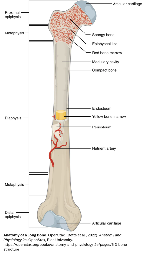
Before diving into 3D exploration, take a close look at the labeled image. It highlights the key regions of a long bone. A typical long bone has five zones: the diaphysis, two metaphyses, and two epiphyses as described below.
- The diaphysis: This is the long, tubular shaft that stretches between the proximal and distal bone ends. Its walls are made of dense compact bone, built to withstand the stress of everyday movement.
- The epiphysis: The wider section at each end of the bone is called the epiphysis (plural = epiphyses), which is filled with spongy bone. Red marrow fills the spaces in the spongy bone and is the site of developing blood cells (red and white) and platelets. The ends of epiphyses are covered with articular cartilage, a thin layer of hyaline cartilage that reduces friction and acts as a shock absorber to form joints with other bones. The epiphysis extends from the end of the bone to the epiphyseal plate (growth plate in children) or epiphyseal line (in adults).
- Metaphysis: This is the area where growth occurs in children, containing the growth plate (epiphyseal plate). It is a highly metabolic and dynamic region that contains a diverse population of cells involved in bone growth and blood formation. It contains spongy bone and red bone marrow.
- The metaphyses contain the epiphyseal plate, which is a layer of hyaline (transparent) cartilage in a growing bone. When the bone stops growing in early adulthood (approximately 18–21 years), the cartilage is replaced by osseous tissue and the epiphyseal plate becomes an epiphyseal line.
- In adults, it transfers loads from weight-bearing joint surfaces to the diaphysis.
Types of bone marrow
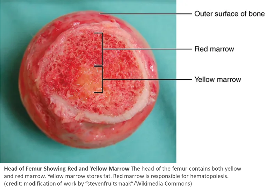
- Red marrow: The primary site for hematopoiesis, or the production of all blood cells. In adults, it is found in spongy bone tissue, such as the heads of the femur and humerus.
- Yellow marrow: Primarily consists of adipose tissue (fat cells) and serves as a storage site for energy in the form of triglycerides. It is found in the medullary cavity of long bones.
Other Key Features
Medullary Cavity
A hollow space within the diaphysis lined by spongy bone and filled with yellow marrow, which stores fat and serves as an energy reserve. It also contains mesenchymal stem cells that can make bone, cartilage or muscle cells. In children, it is almost completely filled with red marrow (red marrow is replaced with yellow marrow with age).
Periosteum
A fibrous membrane covering the outer surface of the bone (except at joint surfaces). It has:
- An outer fibrous layer of dense regular connective tissue for protection.
- An inner osteogenic layer containing bone-forming cells.
It also houses blood vessels, nerves, and lymphatic vessels, and serves as an attachment point for tendons and ligaments.
(Betts et al., 2022)
Endosteum
A delicate membrane lining the medullary cavity, where bone growth, repair, and remodeling occur. (end- = “inside”; oste- = “bone”)
Fun Fact: The femur and tibia are the body’s strongest long bones—they carry most of your weight when you walk, run, or jump! This structural organization allows long bones to be both strong and lightweight—perfectly suited for movement, support, and protection.
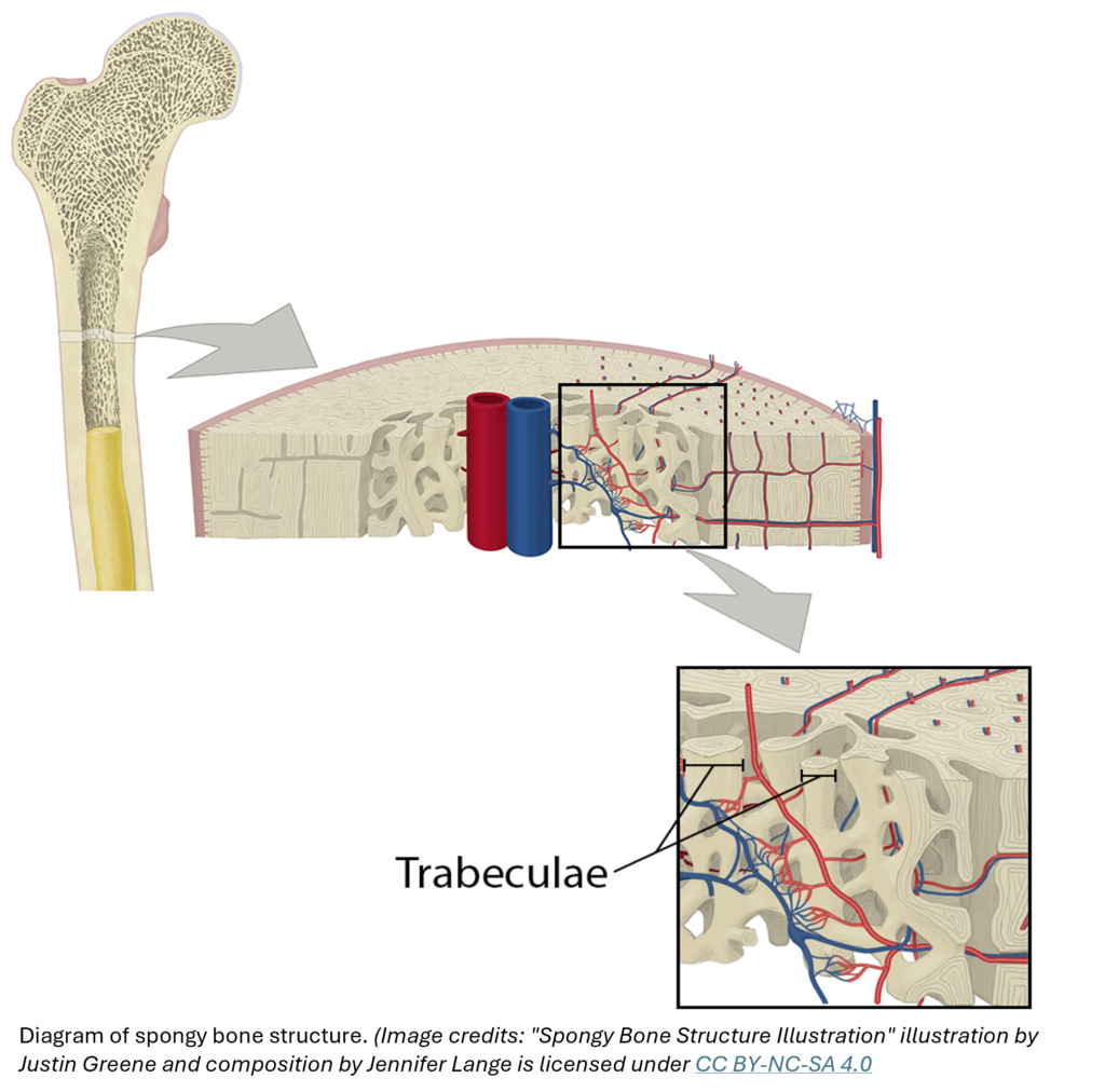
The diaphysis isn’t just a hollow shaft—it’s a powerhouse of structural support and metabolic activity. To understand how it functions, we need to zoom in on the two key types of bone tissue it contains: compact bone and spongy bone (a.k.a. trabecular or cancellous bone).
Use the dropdowns below to explore each type in detail & then reinforce your knowledge by exploring in 3D.
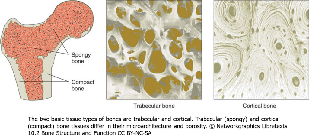
Compact bone is the denser, stronger of the two types of bone tissue. It can be found under the two outer linings in the diaphyses of long bones, where it provides support and protection. It is also found as a thin lining in a portion of the epiphyses.
This cross-sectional view of compact bone below shows the basic structural unit, the osteon (Betts et al. 2022). Each osteon is a cylindrical structure that contains a central canal surrounded by concentric rings of bone matrix—this is the basic unit of compact bone. Take note of the presence of blood vessels, nerves, and lymphatic vessels in each osteon that nourish compact bone. Take note of the presence of spongy bone in the diaphysis as well.
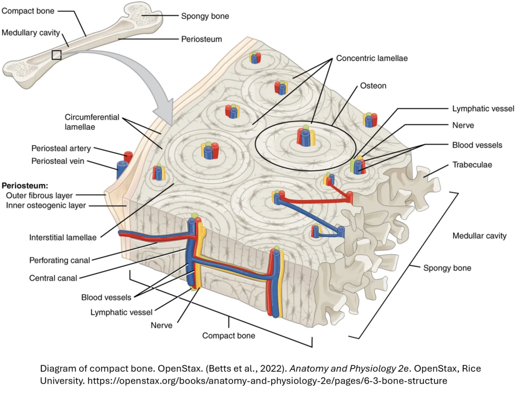
Spongy bone lines the medullary cavity to provide space for the bone marrow. Like compact bone, spongy bone, also known as cancellous bone, contains osteocytes housed in lacunae, arranged in concentric lamellae. Unlike the osteons of compact bone, the sets of lamellae do not surround a hollow central canal and they are not arranged in parallel columns alongside each other, but rather in a lattice-like network of matrix spikes called trabeculae (singular = trabecula) (see figure below). Each trabecula is wrapped in a layer of endosteum. The trabeculae may appear to be a random network, but each trabeculae forms along lines of stress to provide strength to the bone. If the stress on the bone changes, the trabeculae can be quickly remodeled to adapt to the new forces. The spaces of the trabeculated network provide balance to the dense and heavy compact bone by making bones lighter so that muscles can move them more easily. In addition, the spaces in some spongy bones contain red marrow, protected by the trabeculae, where hematopoiesis (producing red blood cells) occurs.
Two bone types in long bone:
Now that you’ve seen labeled images of compact and spongy bone, it’s time to explore these features in 3D.