Learning Objectives
By the end of this section, you will be able to:
- Describe the bones that form the pectoral girdle
- List the functions of the pectoral girdle
Introduction to the Pectoral Girdle
The pectoral girdle plays a vital role in connecting the upper limbs to the axial skeleton, enabling a remarkable range of motion and supporting the dynamic functions of the arms and shoulders. Composed of the scapula (shoulder blade) and clavicle (collar bone), each girdle forms the structural foundation above the arms. As you explore this section, consider how the unique shapes and articulations of these bones contribute to both stability and mobility for the wide variety of movements we rely on every day.
Which bone do you think contributes more to shoulder mobility—clavicle or scapula—and why?
Use your reasoning based on shape, position, and potential movement. Then, explore the dropdowns to test your hypothesis!
Overview:
The clavicle, or collarbone, is a slender, S-shaped bone that acts as a strut between the sternum and scapula. It plays a key role in shoulder mobility and upper limb support.
Key Features:
- Shape: Long, curved like an “S”
- Medial End: Sternal end articulates with the manubrium of the sternum
- Lateral End: Acromial end connects with the acromion process of the scapula
- Function: Supports the shoulder and allows the arm to swing freely from the trunk

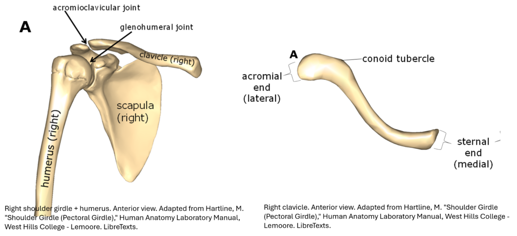
💡 Tip: Use the image to identify the sternal and acromial ends and observe the curvature.
🌐 Explore in 3D:
- 🔗 Sketchfab – Labeled Clavicle Model
View the clavicle in isolation with labeled anatomical features. 💡 Click each numbered item to reveal its name and description—perfect for self-quizzing! - 🔗 AnatomyZone – Clavicle View
Rotate and zoom to explore clavicle landmarks in context with the skeleton. 💡 Tip: Jump to page 5 of 6 to view muscle & ligament attachment sites and their relationships to the bone.
sternal end
acromial end
The clavicle attaches the scapula to the sternum, providing stability and limiting excessive movement. Without this stable connection, the scapula would lose its anchor to the axial skeleton and become much more mobile. This increased mobility could make shoulder movements less controlled and reduce the efficiency of arm motion, while also increasing the risk of injury to surrounding muscles and joints.
The S-shaped curve of the clavicle provides both strength and flexibility. Here’s why it matters:
- Shock Absorption: The curve helps the clavicle absorb and distribute mechanical stress from the arm and shoulder—especially during falls or impacts—reducing the risk of fractures.
- Range of Motion: Its shape and position allow the shoulder to move freely in multiple directions, helping the arm swing away from the trunk.
- Muscle/Ligament Attachment: The curvature creates surfaces for muscle and ligament attachment, supporting shoulder stability and movement (see pages 5/6 in Anatomy Zone).
💡 Think of the clavicle as a flexible bridge—it connects the axial skeleton to the upper limb while allowing dynamic motion and absorbing force.
The coracoid process projects anteriorly from the scapula and serves as an attachment point for:
- Conoid ligament
- Trapezoid ligament
These two ligaments together form the coracoclavicular ligament, which stabilizes the clavicle to the scapula.
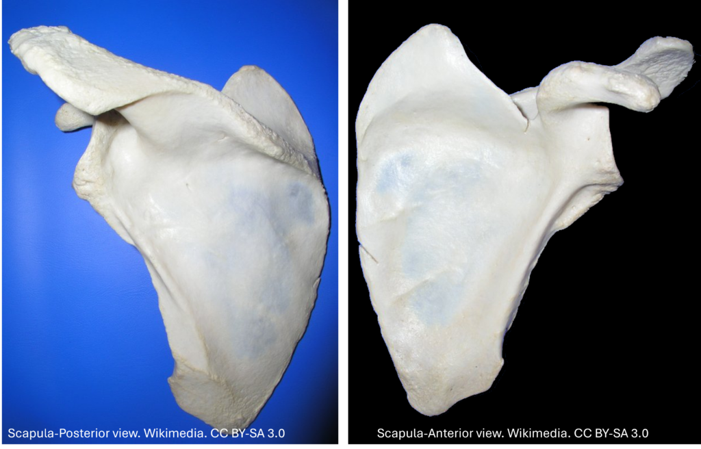
The scapula may be a thin, flat bone—but it plays a powerful role in shoulder movement and stability. In this section, you’ll explore its structure from multiple angles and connect key bony landmarks to muscle attachments and joint function.
Use the activities below to rotate, inspect, and compare the scapula in 3D with real bone views. Then, test your knowledge by identifying features and explaining their roles in shoulder mechanics.
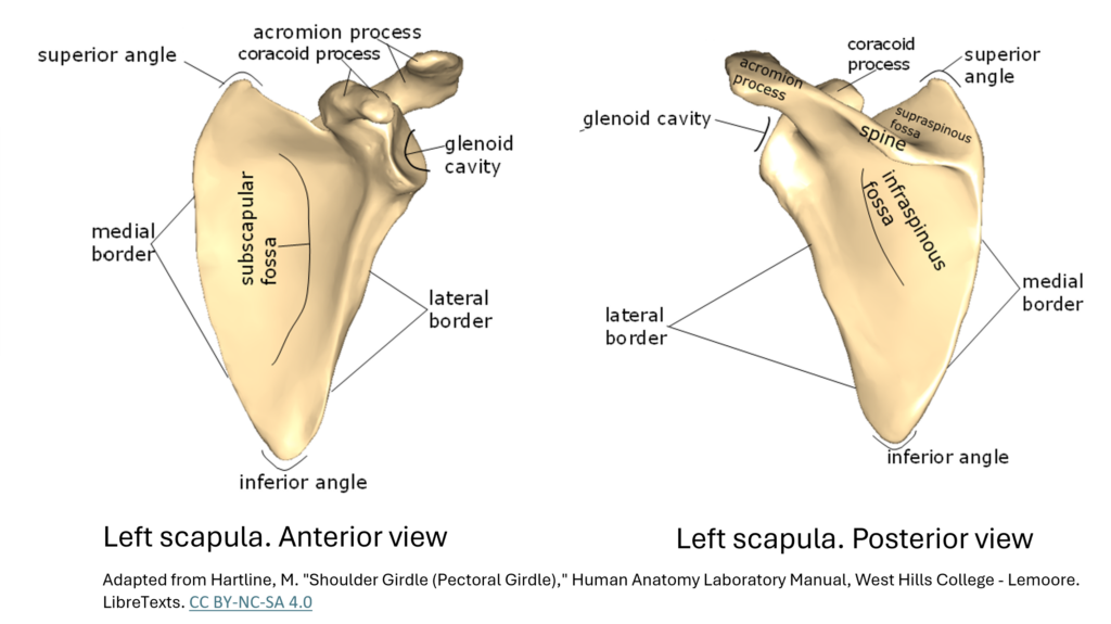
Reference: Key Bony Landmarks
- Glenoid cavity (fossa): Forms the socket of the shoulder joint, articulating with the head of the humerus.
- Acromion process: Extends over the shoulder joint and connects with the clavicle, helping stabilize the joint from above.
- Coracoid process: Serves as an anchor point for ligaments and muscles that support the shoulder.
- Spine of the scapula: Provides attachment for stabilizing muscles like the trapezius and deltoid.
Find these bony landmarks on the images below. Take note of the other labelled aspects of the scapula so you are ready for the ⏱️Beat the Clock Challenge.

- 🔗 Sketchfab – Labeled Scapula Model
View the scapula in isolation with labeled anatomical features. 💡 Click each numbered item to reveal its name and description—perfect for self-quizzing! - 🔗 AnatomyZone – Scapula View
First, rotate and zoom to explore scapula landmarks in context with the skeleton. 💡 Tip: Go through each of the 5 pages to view its unique features in isolation. Remember to click on bones for identification & to take note of anterior and posterior landmarks as you rotate the bone.
Reinforce your recognition of key features by studying anterior and posterior views of the scapula using real bone images. These views help you recognize key features like the spine, acromion, and coracoid process before diving into interactive labeling.
💡 Label these images on your own using the 3D models and descriptions above as a guide. Prepare for the labelling challenges in the Test Your Knowledge section.
Can you identify the spine of the scapula?
What is its function and which muscles attach here?
Which part of the scapula forms the socket for the shoulder joint?
(Hint: It articulates with the head of the humerus.)
Locate the acromion and coracoid process.
How do these projections differ in shape and function?
Which features of the scapula help stabilize the shoulder joint?
Think about both bony landmarks and muscle attachments.

Test Your Knowledge
Answer:
The scapula serves as the foundation for shoulder movement, providing attachment points for muscles and forming part of the shoulder joint. A fracture could limit the stability of the glenoid cavity and disrupt muscle attachments, reducing the range of motion and strength in the upper limb.
Reflect & Reinforce:
Which scapular landmark do you think plays the biggest role in stabilizing the shoulder joint—and why?
The glenoid cavity is the most critical landmark for stabilizing the shoulder joint because it forms the socket of the ball-and-socket articulation with the humeral head. This connection allows for a wide range of motion while maintaining joint integrity. Although the glenoid cavity is relatively shallow, stability is enhanced by surrounding structures like the labrum (fibrocartilaginous ring) and rotator cuff muscles. Without this landmark, the shoulder would lose its primary articulation point, severely limiting mobility and strength (Chang et al., 2025).
Other landmarks, such as the acromion and coracoid process, also contribute by serving as attachment sites for ligaments and muscles that reinforce the joint.
Go to the purpose games site & press play. Use the figure provided in this section to label the scapula. See how fast you can get!
Betts, J. G., Young, K. A., Wise, J. A., Johnson, E., Poe, B., Kruse, D. H., Korol, O., Johnson, J. E., Womble, M., & DeSaix, P. (2022). Anatomy and Physiology 2e. OpenStax. https://openstax.org/books/anatomy-and-physiology-2e/pages/8-1-the-pectoral-girdle
Chang LR, Anand P, Varacallo MA. Anatomy, Shoulder and Upper Limb, Glenohumeral Joint. [Updated 2025 Mar 3]. In: StatPearls [Internet]. Treasure Island (FL): StatPearls Publishing; 2025 Jan-. Available from: https://www.ncbi.nlm.nih.gov/books/NBK537018/
Hartline, M. (n.d.). Shoulder Girdle (Pectoral Girdle). In Human Anatomy Laboratory Manual. West Hills College – Lemoore. Retrieved from https://bio.libretexts.org/Courses/West_Hills_College_-Lemoore/Human_Anatomy_Laboratory_Manual(Hartline)/09%3A_Appendicular_Skeleton/9.02%3A_Shoulder_Girdle_Pectoral_Girdle
Lumen Learning. (n.d.). 6.01: The Pectoral girdles. In Human Anatomy Laboratory Manual. Medicine LibreTexts. Retrieved from https://med.libretexts.org/Bookshelves/Anatomy_and_Physiology/Human_Anatomy_Laboratory_Manual/06%3A_The_Appendicular_Skeleton/6.01%3A_The_Pectoral_girdles