Learning Objectives
By the end of this section, you will be able to:
- Define the pelvic girdle and describe the bones of the pelvis
- Explain the three regions of the hip bone and identify their bony landmarks
- Distinguish between the male and female pelves (plural for pelvis).
Understanding the Pelvic Girdle vs. Pelvis
Before exploring the anatomy, it’s important to distinguish these two terms:
- The pelvic girdle refers specifically to the two hip bones (coxal bones) that attach the lower limbs to the axial skeleton.
- The pelvis is a broader structure that includes the pelvic girdle plus the sacrum and coccyx.
This distinction matters because the pelvic girdle provides limb attachment and weight transfer, while the entire pelvis forms the basin-like cavity that supports abdominal organs and anchors muscles.
Use the table & figure provided in the dropdown below to compare the pelvic girdle and pelvis. Identify the components of each and then answer the check-in question to reinforce your understanding.
Pelvic Girdle vs. Pelvis Comparison Table
This table outlines the key differences between the pelvic girdle and the pelvis, including their components and functions.
| Structure | Components | Function |
| Pelvic Girdle | Two hip bones (coxal bones) | Attaches lower limbs to axial skeleton; transfers weight |
| Pelvis | Pelvic girdle + sacrum + coccyx | Forms the basin-like cavity; supports organs; anchors muscles |
Pelvic Girdle
The Pelvic Girdle. Wikimedia. (CC BY 2.1 JP)
Pelvis
The Pelvis. Wikimedia. (CC BY 2.1 JP)
The Hip Bone: Structure & Landmarks
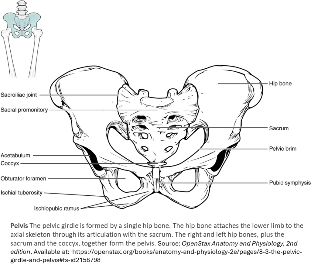
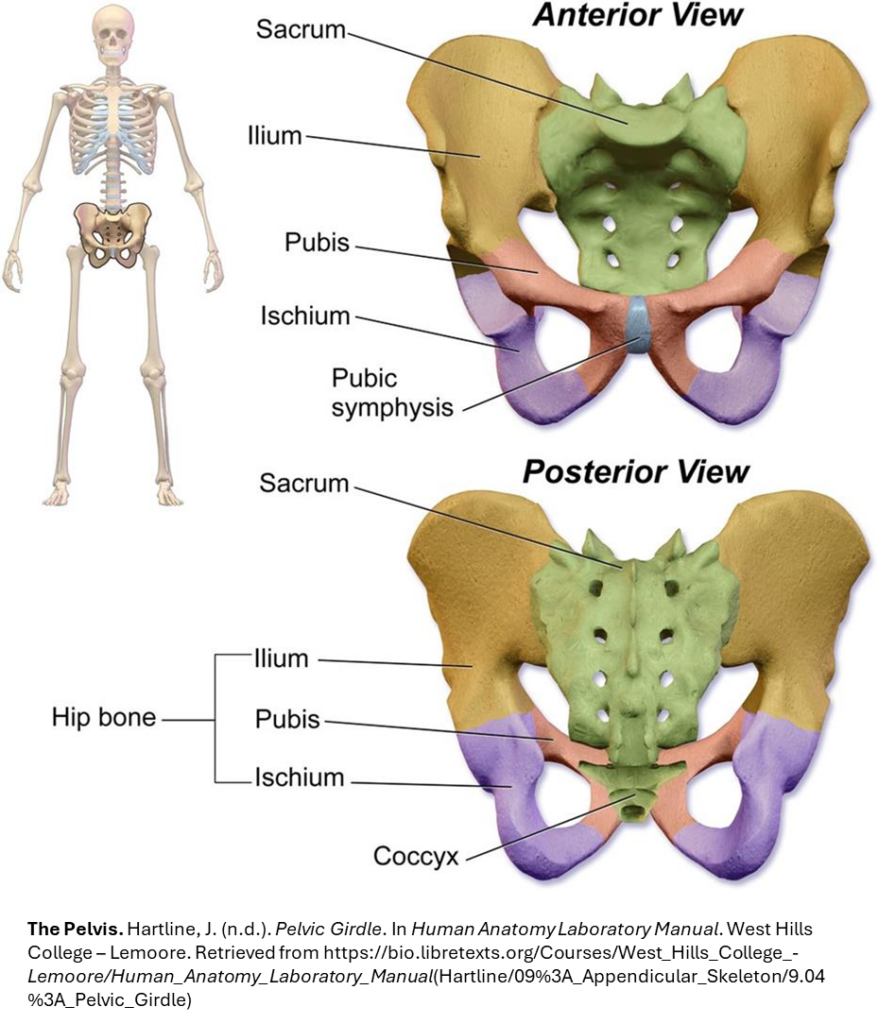
The hip bone, or coxal bone, forms the pelvic girdle portion of the pelvis. The paired hip bones are the large, curved bones that form the lateral and anterior aspects of the pelvis. At birth, each coxal bone starts out as three separate bones – the ilium, (ILL-ee-um), the ischium, (ISH-ee-um) and the pubis (PYOO-bus) bones – joined by hyaline cartilage. By the age of 25, these three bones have fully fused into a single coxal bone. We still subdivide the fully-formed coxal bone into three regions based on the positions of the three bones that fused to form it, each region named after the bone that gave rise to that region. Identify the hip bones on the figure provided in the dropdown below.
On the anterior side of the pelvis, the pubis portions of the two coxal bones do not articulate with each other, but instead are joined with a small piece of cartilage called the pubic symphysis (“PYOO-bick SIM-fiss-is”).
- Ilium: In a fully fused coxal bone, the ilium is the most superior portion, forming the “wing” that makes up the most prominent part of the hip bone. The ilium is where the sacrum attaches to each coxal bone to complete the pelvic bowl.
- Ischium: Positioned posterior to the pubis in anatomical position. You “sit on the ischium,” which is thicker and stronger than the pubis, allowing it to support body weight.
- Pubis: Forms the anterior portion of the hip bone. The pubis curves medially and joins the opposite pubis at the pubic symphysis, a specialized joint.
🔍 Locate these landmarks on the figure.
Can you identify the ilium, ischium, and pubis on the anterior & posterior views of the pelvis?

🧍♀️🧍♂️ Comparing Male and Female Pelves
The pelvis is one of the few bones that can help determine biological sex in skeletal remains. Differences between male and female pelvises reflect their distinct functions—especially the female pelvis’s role in childbirth.
Regions of the Pelvic Cavity
The space enclosed by the bony pelvis is divided into two regions:
1. Greater Pelvis (False Pelvis)
- Location: Broad, superior region above the pelvic brim
- Boundaries: Defined laterally by the large, fan-like portion of the upper hip bone
- Contents: Portions of the small and large intestines
- Association: Closely related to the abdominal cavity → called the false pelvis
2. Lesser Pelvis (True Pelvis)
- Location: Narrow, rounded space below the pelvic brim
- Contents: Bladder and other pelvic organs
- Association: Called the true pelvis because it houses pelvic organs
Pelvic Brim (Pelvic Inlet)
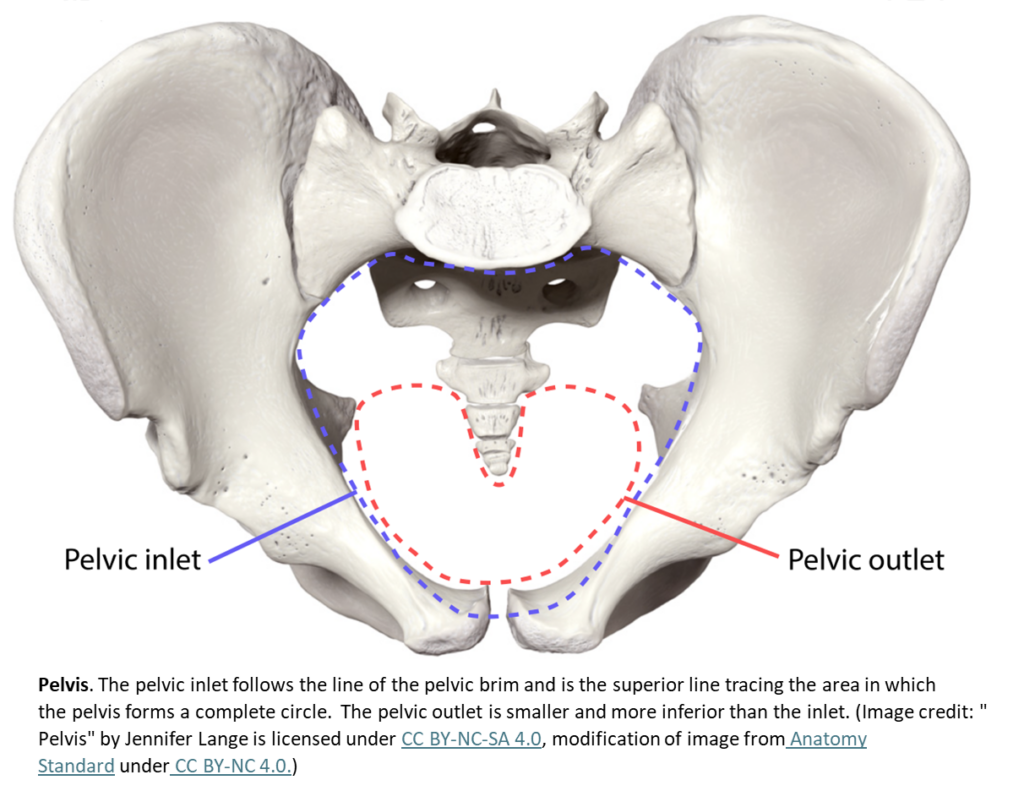
- Function: Separates the greater pelvis from the lesser pelvis
- Defined by:
- Upper margin of the pubic symphysis (anterior)
- Pectineal line of the pubis
- Arcuate line of the ilium
- Sacral promontory (anterior margin of the superior sacrum)
Note: The pelvic brim is the bony rim that separates the false (greater) from the true (lesser) pelvis. It is the edge of the pelvic inlet.
Pelvic Outlet
- Function: Inferior limit of the lesser pelvic cavity
- Defined by:
- Inferior margin of the pubic symphysis (anterior)
- Ischiopubic ramus, ischial tuberosity, sacrotuberous ligament, and tip of the coccyx (posterior)
Orientation: Because of the anterior tilt of the pelvis, the lesser pelvis is angled from anterosuperior (pelvic inlet) to posteroinferior (pelvic outlet).
One of the most reliable ways to determine biological sex from a skeleton is by examining the pelvis. Why? Because its structure reflects functional differences—especially the female pelvis’s role in childbirth.
🌐 Click here to compare unlabelled male/female pelves on Sketchfab in 3D!
🌐 Click here to compare male/female pelves with some labelling in 3D!
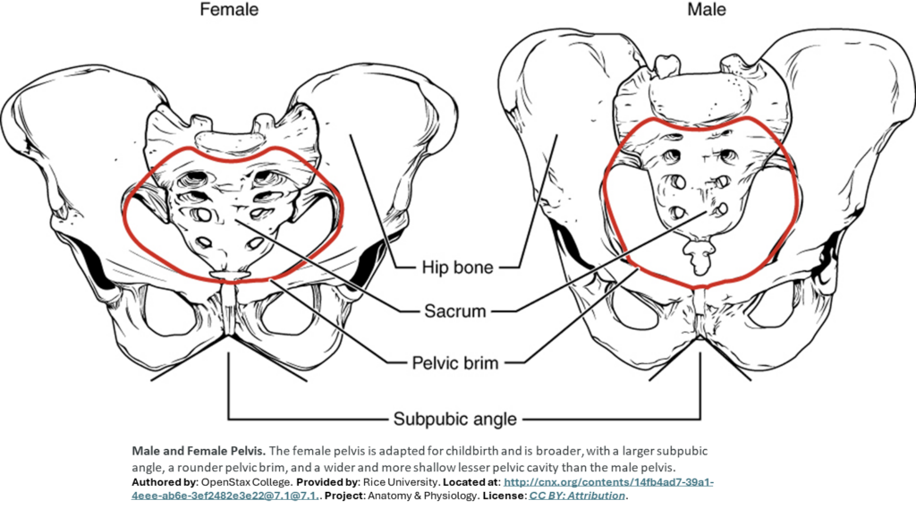
The female pelvis can be distinguished from the male pelvis by a number or criterion, three of which are shown in the figures below and others are summarized in the table.
🔍 Look for These Features in the Figures Below:
Summary Table of Differences
| Feature | Female Pelvis | Male Pelvis |
| Pelvic Weight | Bones are lighter and thinner | Bones are thicker and heavier |
| Pelvic Inlet Shape | Round or oval | Heart-shaped |
| Lesser Pelvic Cavity | Shorter and wider | Longer and narrower |
| Subpubic Angle (=pubic arch) | Greater than 90° | Less than 90° |
| Obturator foramen | Oval | Round |
| Pelvic Outlet Shape | Rounded and larger | Smaller |
| Sacrum | Short, wide, rounder, less inwardly curved | Long and narrow |
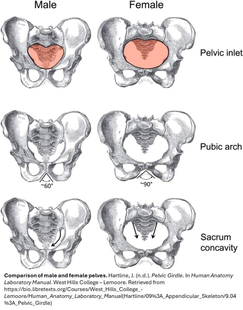
The feature most directly related to childbirth is the pelvic inlet shape (and by extension, the overall width of the lesser pelvic cavity). See pelvic inlet circled in the diagram below.
Why?
- A wider, round or oval pelvic inlet in females provides space for the fetus to pass through during delivery.
- This adaptation also influences other features like the subpubic angle and pelvic outlet, which together create a birth canal that is larger and less obstructed compared to the male pelvis.